Graham S. Devereux
- Professor of Respiratory Medicine
- Division of Applied Health Sciences
- University of Aberdeen
- Consultant in Respiratory Medicine
- Aberdeen Royal Infirmary
- Aberdeen, UK
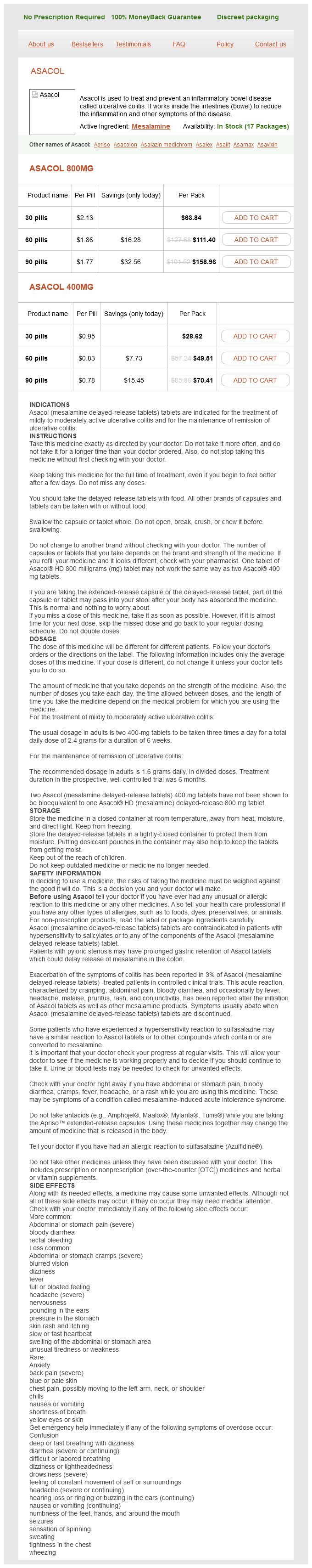
Mesalamine dosages: 800 mg, 400 mg
Mesalamine packs: 30 pills, 60 pills, 90 pills
Discount 800 mg mesalamine visa
Endocarditis is a relatively unusual reason for stroke, however stroke in sufferers with endocarditis is pretty common, clinically apparent in over 1/3 of sufferers however asymptomatically current in one other 50%. Larger vegetations, ones on the mitral and to less extent aortic valve, and ones from Staphylococcus aureus carry higher risks. Smaller or streptococcal vegetations still can cause emboli, as can vegetations in the best heart. Likewise, antiplatelet agents are unlikely to confer additional profit but will increase hemorrhagic threat (albeit less than anticoagulation). Risk with anticoagulation is additional elevated by the potential of the rare complication of a mycotic aneurysm-arterial wall erosion and dilatations from invasion of infective agent. Additionally, in sufferers with embolization (whether cranial or systemic), screening for occult embolization after initiation of antimicrobial remedy could be useful. Recurrent infarction several days after the initiation of appropriate antimicrobial therapy is an indication for valvular surgical procedure as it suggests medical failure. Cerebral hemorrhage or vascular imaging that recommend aneurysm formation requires a formal angiogram for prognosis and potential treatment of a mycotic aneurysm. Recurrent embolization after initiation of acceptable antimicrobial therapy is an indication for consideration of valvular surgery. Infective endocarditis because of Staphylococcus aureus: deleterious effect of anticoagulant remedy. She had coronary angiography that revealed extreme 3-vessel coronary artery disease. Before the process the surgeon famous a left carotid bruit and ordered an ultrasound examination of the neck arteries. The Doppler examination was in maintaining with 60% stenosis of the left inside carotid artery and fewer than 30% stenosis of the best. Approximately 30�40% of these are detected within the instant postoperative period, while the remaining are discovered after the patient awakens from anesthesia uneventfully. The mechanism of the majority of perioperative strokes is embolism from the guts or aorta. Risk components for perioperative stroke embody advanced age, history of hypertension, congestive coronary heart failure, diabetes, and peripheral vascular illness. Aortic atherosclerosis is a serious danger for perioperative stroke and encephalopathy. It is essential to identify patients with symptomatic disease and those in whom carotid artery illness has triggered strokes shown by brain imaging. Patients with asymptomatic carotid artery stenosis are sometimes recognized preoperatively by auscultation of a carotid bruit. Carotid bruits are associated with long-term threat of stroke during the following years. Often the stroke is in a territory incongruent with the affected vessel, suggesting that the presence of a bruit is a marker for atherosclerosis and stroke danger normally. This discrepancy is essentially as a result of the varying nature of the studies that have addressed this concern, and so uncertainty as to the significance of asymptomatic carotid stenosis stays. Time is better directed towards evaluating sufferers for aortic atherosclerostic and poor cardiac function, as these are more frequent sources of embolism. The most common mechanism of perioperative stroke in sufferers with carotid stenosis is embolism from the guts or aorta. Perioperative stroke is most often acknowledged after a delay from anesthesia restoration. Continuing controversy in the administration of concomitant coronary and carotid disease: an outline. A systematic evaluation of outcomes in sufferers with staged carotid artery stenting and coronary artery bypass graft surgical procedure. Determination of etiologic mechanisms of strokes secondary to coronary artery bypass graft surgery. Type 1A diabetes usually presents with acute symptomatic hyperglycemia and diabetic ketoacidosis.
Generic mesalamine 800 mg buy on line
Deep respiration, tightening and stress-free your muscular tissues, stretching, and leg lifts are possible. Walking is an excellent form of cardio train that may assist you to keep fitness and stamina. This applies to both the patient awaiting transplantation and the recipient of a transplanted organ. Even though the extent of exercise could also be gentle, it is very important heat up to begin with and to calm down at the end. Stay nicely hydrated, albeit inside the limits your doctor has really helpful when you have fluid retention problems. Also, try to vary your exercises to maintain them fascinating, as a end result of your degree of fitness has a direct correlation with your recovery after transplantation. In many ailments, adhering to a specific food regimen is helpful in controlling the development of the illness. For example, sufferers with heart illness can scale back the chance of heart attack by following a low-fat, low-cholesterol diet. Instead, you must concentrate on eating a usually healthy diet with the really helpful stability of meals groups. Eating a wholesome quantity of fruits, vegetables, cereals, and meat offers you with the correct steadiness of carbohydrates, fat, and proteins. Some sufferers with liver disease have diminished appetites and require supplementation with small-volume, high-calorie, wellbalanced liquid meals similar to Ensure, Boost, and Sustacal. For example, patients with fluid retention (ascites or edema) could also be on a low-sodium (approximately 2,000 milligrams per day) and fluidrestricted (approximately 2 liters or sixty seven ounces per day) food regimen. A dietitian from the liver transplant staff can provide you practical advice on tips on how to meet these objectives. Patients with hepatic encephalopathy are often advised to significantly cut back their protein intake to achieve better management of their encephalopathy. This advice can be harmful and should result in extreme protein malnutrition, which in turn may lead to muscle losing, weakness, and poor wound healing. Instead, the calories and energy brought in by their meals consumption are used to address the needs of the liver and different organs. In fact, these sufferers need extra energy and proteins than wholesome people just to keep their weight and muscle mass. Thus any limitation of protein results in progressive loss of muscle and body weight. Vitamin supplementation may also be necessary, particularly in patients with the cholestatic liver diseases of main biliary cirrhosis and first sclerosing cholangitis. If sufferers with liver illness can eat a nutritious diet, then adding the standard multivitamins to the food regimen is often not needed. Almost all oral medications are absorbed in to the bloodstream and carried immediately to the liver for processing. Most prescription medicines are secure for the liver, though some require a discount in dose for patients with liver disease. Several generally prescribed medicines are worthy of specific point out at the facet of liver disease. Acetaminophen is usually combined with other ache medicines similar to oxycodone (Percocet), hydromorphone (Vicodin), and Darvocet. Contrary to well-liked belief, acetaminophen can be taken safely by sufferers with liver disease, as long as they adhere to some limitations. Fortunately, a detoxifier, known as glutathione, is waiting for the toxin to arrive in the liver. The damaged liver may have a slower price of glutathione production however nonetheless has a replenishable supply. Because acetaminophen is found in lots of widespread medicines, you must acknowledge that the whole every day dose might come from completely different sources of acetaminophen. Cholesterol-Lowering Agents Cholesterol management has improved dramatically because the introduction of the cholesterol-lowering agents known as statins. One of the unwanted effects of this class of medicine is liver cell toxicity, though this downside occurs in solely a minority of sufferers.
Buy cheap mesalamine
Other cardiac (valvular atresia or stenosis or coarctation of aorta) or noncardiac (heterotaxia, cystic hygroma, congenital diaphragmatic hernia) anomalies are regularly associated. Chromosomal anomalies could also be additionally associated (trisomy 18, Klinefelter syndrome, DiGeorge syndrome, or Goldenhar syndrome). The connection between each atria and one ventricle may be by way of one or two atrioventricular valves. Although a rudimentary accessory ventricle may also exist, only one ventricle is useful receiving the circulate from each atria. Etiology and Pathophysiology the morphologic spectrum of double-inlet single ventricle is broad, with quite lots of segmental patterns and anatomic variables, together with differences in anatomic sample of the one ventricle (left, proper, or indeterminate); the sizes of the atrioventricular valves or atresia of one atrioventricular valve; variable ventricular looping and ventriculoarterial relationship; and obstruction to systemic or pulmonary outflow (or each systemic and pulmonary outflow). The most typical kind is a single left ventricle with a rudimentary proper ventricle, two atrioventricular valves, and aorta disposed anteriorly and to the left of the pulmonary artery. Outflows from the nice arteries are usually anomalous with usually an anterior and left aortic disposition. Anatomic, imaging, and medical traits of double-inlet, double-outlet right ventricle. Mid-term results for double inlet left ventricle and comparable morphologies: timing of Damus-KayeStansel. The terms heterotaxy syndrome, cardiosplenic syndrome, right and left isomerism, and situs ambiguus are sometimes used to describe these defects. It is related to a really poor prognosis owing to high in utero and neonatal mortality. Heart block, atrioventricular septal defect, double-outlet proper ventricle, proper or left ventricular outflow tract obstruction, total anomalous pulmonary vein drainage, persistence of left superior vena cava, and aorta and inferior vena cava positioned on the same belly side may be present. However, associations with chromosome 22q11 deletion (DiGeorge syndrome), trisomy 18, trisomy thirteen, and different chromosomal deletions or inversions have been described. Complete coronary heart block, complicated cardiac abnormalities, and fetal hydrops are poor prognostic options. Right isomerism is related to a worse prognosis with a really high mortality rate (>90%) within the 1st yr after delivery. Atrial isomerism also can be recognized within the first trimester as increased nuchal translucency, cardiac rhythm disturbance, or hydrops. A proper situs belly exploration is important to detect typical anomalies corresponding to abdomen dextroposition, intestinal obstruction, and inferior vena cava malposition (Video 92-2). Other typical anomalies, corresponding to asplenia or pulmonary lobulation, could require postnatal imaging or necropsy. Etiology and Pathophysiology Atrial isomerism manifests with a diversified spectrum of cardiac malformations with viscerocardiac heterotaxy occurring generally. Typical options include asplenia, bilateral trilobed lungs, intestinal malrotation, irregular drainage of the pulmonary veins, persistence of the left superior vena cava, aorta and inferior vena cava positioned on the same abdominal aspect, atrioventricular septal defect, and ductus-dependent pulmonary move. Fetal anomalies embody bilateral morphologic left atrial appendages, bilobed lungs, a number of splenuli, and malrotation of the intestines. The nomenclature, definition and classification of cardiac buildings in the setting of heterotaxy. Clinical options, administration and end result of youngsters with fetal and postnatal diagnoses of isomerism syndromes. The syndrome of left isomerism: sonographic findings and end result in prenatally identified instances. Spectrum of heart problems: accuracy of analysis and end result in fetal heterotaxy syndrome. With the advance in long-term outlook for these patients on account of modern cardiac surgery, intraabdominal anomalies have turn into increasingly relevant because they may have an result on longterm end result. Immediate and rising hypoxemia and acidosis are the rule, which could be reversed solely by emer gency postnatal cardiac surgical procedure. The four pulmonary veins drain in to a venous collector positioned behind the left atrium, from which a vertical vein runs up to attain the innominate vein, and this terminates in the best superior vena cava and proper atrium. It has been reported that the vertical vein may be obstructed, however this is rarely extreme in the perinatal period. The pulmonary veins drain through the coronary sinus or immediately in to the best atrium. The pulmonary veins converge in to a descending vertical vein that reaches the stomach by way of the diaphragm.
Order mesalamine 800 mg line
Common indications Dextromethorphan is indicated for the momentary aid of nonproductive cough. Common opposed effects Common opposed effects are gentle and infrequent and may embody dizziness, fatigue, gastrointestinal disturbances, and drowsiness. Dextromethorphan has been related to serotonergic results and at higher doses may trigger confusion, nervousness, restlessness, dysarthria, irritability, nausea and vomiting. Some dextromethorphan-containing merchandise contain tartrazine, which may cause allergic-type reactions (including bronchial asthma symptoms) in sufferers with a selected sensitivity (Ardern & Ram, 2001). Although the overall prevalence of tartrazine sensitivity within the basic inhabitants is low, it may happen in sufferers who also have aspirin hypersensitivity, though this was disputed in a recent research evaluating tartrazine sensitivity in sufferers with nonsteroid anti-inflammatory drug sensitivity (Pestana, Moreira, & Olej, 2010). Beginning in 2007, the Centers for Disease Control issued warnings for the chance of great injury and fatal overdose from cough and chilly merchandise administered to kids less than 2 years old. Subsequently, the Food and Drug Administration Nonprescription Drug Advisory Committee and Pediatric Advisory Committee recommended that merchandise containing dextromethorphan, pseudoephedrine, chlorpheniramine, diphenydramine, brompheniramine, phenylephrine, clemastine, and guaifenesin not be used in kids less than 6 years old. The Food and Drug Administration issued a Public Health Advisory recommending that nonprescription merchandise (though prescription merchandise also containing these components could be included) not be utilized in youngsters underneath 2 years old. Principles of lung therapeutics sixty seven Generic and model names There are numerous brand and generic merchandise obtainable without prescription. Formulations Dextromethorphan is on the market in liquid, tablet, softgel, and dissolving movie formulations. Codeine Mechanism of motion Codeine is a narcotic opioid that acts centrally within the cough heart within the medulla by binding primarily to �-opioid receptors and possibly -opioid receptors (Takahama & Shirasaki, 2007). Codeine also has a drying impact on the mucosa of the respiratory tract and will increase the viscosity of bronchial secretions. Common indications Codeine is indicated for the short-term reduction of nonproductive cough. Common adverse results probably the most severe opposed effects include respiratory depression and arrest, circulatory melancholy, shock, and cardiac arrest. Codeine can also cause drowsiness, dizziness, confusion, insomnia, nervousness, dysphoria, euphoria, mood alterations, and anxiety. Gastrointestinal effects may include anorexia, nausea, vomiting, and constipation. Anticholinergic effects are infrequent but can embrace sinus bradycardia, sinus tachycardia, changes in blood pressure, and syncope, as properly as dry mouth, blurred imaginative and prescient, or urinary retention. Numerous model and generic products can be found and distributed at retail without sixty eight Nursing Care in Pediatric Respiratory Disease prescription, but only by a registered pharmacist; in some states, a prescription could additionally be required. Formulations Products can be found as tablets and oral liquids alone or together with expectorants and decongestants. Diphenydramine, promethazine, and other first-generation antihistamines Mechanism of action First-generation antihistamines similar to diphenhydramine and promethazine have direct suppressive actions on the cough middle in the medulla. Common indications First-generation antihistamines can be used for the momentary reduction of nonproductive cough. Common antagonistic results probably the most frequent opposed results include drowsiness, dizziness, and xerostomia. Central nervous stimulation is more likely to happen in children, and effects can include agitation, insomnia, elevated urge for food, restlessness, palpitations, muscle spasms, and seizures. Anticholinergic effects can embrace xerostomia, insomnia, urinary retention, nervousness, mydriasis, xerophthalmia, and blurred vision. Quinidine-like anesthetic results can embrace sinus tachycardia and cardiac arrhythmias. Ciliary motion is improved with the flow of less viscous secretions converting a dry, nonproductive cough to a productive cough (Woo, 2008). Common indications Guaifenesin is indicated for loosening and thinning phlegm related to coughs from colds and minor higher respiratory tract infections so as to facilitate clearing of bronchial passages and increasing productive cough. Guaifenesin is also found in quite a few combination merchandise with antihistamines, decongestants, dextromethorphan, and different cough and cold merchandise. Stimulation of postcapillary 1-adrenergic receptors causes vasoconstriction of postcapillary venules in the nasal mucosa (Johnson & Hricik, 1993). These postcapillary venules are capacitance vessels and can accommodate a comparatively large amount of blood. Congestion is caused by increased blood volume, which will increase the volume of the nasal mucosa.
Generic mesalamine 400 mg without a prescription
It could be the consequence of a vascular accident, different ischemic insults, an infection, maternal exposure to toxins, or dying of a cotwin in a monochorionic being pregnant. It is normally recognized at midgestation, nevertheless it also can happen later in pregnancy. The prognosis may be very poor, and termination of being pregnant should be considered if attainable. The clefts are sometimes bilateral and symmetric, offering communication between the lateral ventricles and the external subarachnoid space. Distinguishing a big porencephalic cyst within the area of the sylvian fissure from schizencephaly may be impossible; nevertheless, large porencephalic cysts are usually unilateral with jagged contours. It is probably not the end result of a destructive means of the fetal mind, however somewhat a morphogenetic dysfunction. According to probably the most accepted principle, the cerebral clefts are the results of an alteration in neuronal migration from the germinal matrix. Manifestations of Disease Clinical Presentation the neuropathologic options of schizencephaly embrace (1) hemispheric clefts, lined with pia-ependyma, often bilateral, within the space of the sylvian fissure; (2) communication of the subarachnoid house with the lateral ventricle, with infolding of grey matter alongside the cleft; and (3) multiple related intracranial malformations (polymicrogyria, grey matter heterotopias, absent septum pellucidum, optic nerve hypoplasia, and agenesis of the corpus callosum). The prognosis is often poor; youngsters usually have varied levels of psychological retardation and developmental delay, seizures, and motor abnormalities; not often, asymptomatic instances with type I even have been reported. Glioependymal cysts are often located in the midline and are regularly associated with agenesis of the corpus callosum. Differentiation between arachnoid and glioependymal cysts is impossible prenatally, though the latter condition is extra doubtless when the lesion is in the midline. Arachnoid cysts are the results of an accumulation of cerebrospinal-like fluid between the cerebral meninges. Synonyms for glioependymal cysts include ependymal cysts, neuroepithelial cysts, epithelial cysts, choroidal epithelial cysts, and interhemispheric cysts (occasionally related to agenesis of the corpus callosum). Etiology and Pathophysiology From exterior to internal, the meninges are composed of the dura mater, arachnoid, and pia mater. Arachnoid cysts originate from a fluid assortment between the dura mater and the arachnoid, between the two layers of the arachnoid (intraarachnoid cysts), or between the arachnoid and the pia mater (subarachnoid cysts). They could be major (maldevelopment of the meninges) or secondary (result of trauma, hemorrhage, or infection). When a midline cyst is recognized, it could be very important rule out related agenesis of the corpus callosum. A posterior fossa arachnoid cyst is often difficult to differentiate from primary cerebellar anomalies similar to Dandy-Walker malformation (see Chapter 38). Rare forms embody tumors related to embryonal remnant tissues (craniopharyngiomas) and tumors of ependymal origin (glioblastoma multiforme, choroid plexus papillomas). Prevalence and Epidemiology Fetal intracranial tumors are uncommon, with an estimated incidence of 0. They are asymptomatic typically, though they could cause epilepsy, gentle motor or sensory abnormalities, and hydrocephalus in children. A worse end result could additionally be anticipated in case of related obstructive hydrocephalus or agenesis of the corpus callosum. In an animal model, brain tumors have been produced by means of chemical or viral teratogens. Manifestations of Disease Clinical Presentation Teratomas may comprise well-differentiated structures (hair, bone) or undifferentiated tissues with a better tendency toward malignancy. They are usually observed within the pineal region, the suprasellar area, or the fourth ventricle, with mass impact and distortion of the encircling brain structure. Epidermoid tumors derive from epithelial cells and appear as cystic lesions containing desquamation of the inner epithelial lining. They are largely positioned within the suprasellar area, in the temporal lobe, or at the level of the cerebellopontine angle. Germinomas originate from germ cells and are stable lesions occurring within the pineal and suprasellar area. Medulloblastomas appear as friable masses with inner necrosis, growing in the posterior fossa. Craniopharyngiomas derive from remnants of the craniopharyngeal duct and are solid-cystic lesions often positioned within the suprasellar region.
Bignonia sempervirens (Gelsemium). Mesalamine.
- How does Gelsemium work?
- What is Gelsemium?
- Asthma, pain due to migraine headaches, pain due to a condition of facial nerves called trigeminal neuralgia, and other uses.
- Dosing considerations for Gelsemium.
- Are there safety concerns?
Source: http://www.rxlist.com/script/main/art.asp?articlekey=96464
Mesalamine 400 mg buy line
Children with attainable fusions should have radiographs taken periodically as they mature as a result of incomplete vertebral ossification could make fusions troublesome to appreciate in younger youngsters. Tourette syndrome and KlippelFeil anomaly in a toddler with chromosome 22q11 duplication. Klippel-Feil syndrome with congenital conductive deafness: report of case and review of literature. Anomalies of the palms are difficult to diagnose, and malformations range from refined deformities, similar to isolated clinodactyly, to full absence of extremities, similar to phocomelia. Anomalies of the digits could also be isolated or related to other skeletal abnormalities. The differential prognosis for irregular fetal arms is vast and includes chromosomal abnormalities and genetic syndromes. Any of those abnormalities can happen in isolation, or they can be related to a multitude of aneuploidies or genetic syndromes. Two diagnoses that ought to be strongly thought-about within the case of an absent or irregular (hypoplastic or triphalangeal) thumb are Fanconi anemia and Holt-Oram syndrome. The most common higher extremity malformations involve the radial, thenar, and carpal bones, with the left side often extra severely affected than the proper. Cardiac manifestations embody atrial septal defect, ventricular septal defect, and conduction abnormalities. Congenital heart malformations are current in 75% of people with Holt-Oram syndrome. These breakpoints can be misrepaired as chromatid interchanges, further contributing to chromosome damage. In terms of prenatal prognosis, besides abnormal thumbs, abnormalities of the cardiac, gastrointestinal, and renal systems can be associated with Fanconi anemia. Prevalence and Epidemiology Global prevalence of Fanconi anemia is estimated to be 1: 1 million to 5: 1 million, affecting women and men and all races and ethnic teams. It is an autosomal recessive dysfunction, and the heterozygous mutation carrier frequency is estimated to be between 0. Less generally, supernumerary thumbs, microcephaly, microphthalmos, hypoplastic kidney, double ureter, or anomalies of the center or ear may be current. Hematologic findings usually include pancytopenia with bone marrow failure in the first 10 years of life with the incidence of bone marrow failure 90% by the 4th decade. There is a big threat of growing hematologic malignancies (predominantly acute myelogenous leukemia) and other nonhematologic malignancies of the top and neck, pores and skin, gastrointestinal tract, and genital tract. It is autosomal dominant, with almost 100 percent penetrance and variable expression of the phenotype. First-trimester examinations of the fetal digits may be indicated when early analysis of a fetal genetic syndrome is desired, similar to in a household with a earlier youngster with an autosomal recessive syndrome with related hand abnormalities or if one of the parents manifests a possible dominant disorder. Alternatively, three-dimensional ultrasound with quantity acquisition and manipulation may be used to show the fetal hand anatomy, including the connection between the thumb and different digits. In many cases, parents also wants to be referred to a medical geneticist for examination. Molecular diagnosis by way of invasive testing could also be available for many disorders, and preimplantation analysis may be potential in circumstances the place the mutations have been established in the family. Delivery is normally routine as nicely, with cesarean section reserved for the same old obstetric indications; however, fetuses with skeletal abnormalities are at increased threat of malpresentation or abnormal extremity position, or each, which can require cesarean delivery. Given the broad variations amongst problems associated with irregular hands, care should be individualized. Surgical correction is possible in many hand abnormalities, especially in cases of isolated abnormalities. However, fetuses with irregular hands that are related to different findings may require further assist at delivery, relying on the precise anomalies, so supply should typically occur at a tertiary center to facilitate early consultation with consultants in medical genetics and neonatal subspecialties, which may include pediatric orthopedics, hand surgery, cardiology, and cardiothoracic surgery. The clinical and genetic spectrum of the Holt-Oram syndrome (heart-hand syndrome). The differential diagnosis for irregular fetal arms is vast and contains aneuploidies and genetic syndromes with various modes of inheritance. Correctly identifying a hand anomaly is crucial as a result of certain hand anomalies are strongly suggestive of specific problems and might provide valuable clues to prenatal analysis.
Purchase line mesalamine
Because the left lobe is the smaller of the 2 lobes, it can be used as a living donor organ only in youngsters or very small adults. The bigger proper lobe is needed when the recipient is an average-size or larger adult. A potential donor should first volunteer to donate a portion of his or her liver to a member of the family or somebody with whom he or she shares robust emotional ties. This not only will increase the chance of an enough donor liver measurement but also ensures that the donor is of authorized age to consent to the process. Careful screening checks have to be carried out to evaluate the well being and suitability of the donor. Many elements have to be thought of in this decision, including the medical points talked about previously. As with any surgical process, there are dangers involved in donating part of the liver. Living donors receive basic anesthesia for the operation, and stay donor liver transplantation is considered main surgical procedure. Living donors may develop complications such as infections, bleeding, and even dying. Similar emotions could happen if, after donation, the recipient has an episode of rejection. It is important that potential donors, recipients, and their households be conscious of these points and have adequate help obtainable if any occur. These helps come from the transplant group, psychological well being professionals, and shut family and friends. Although donating part of the liver offers no direct medical benefit for the donor, it has significant benefits for the recipient. The surgery could be scheduled at a time when the recipient is in pretty good physical situation (timing is very important-the recipient ought to neither be too sick nor too well). A reside donor transplant shortens the length of time the recipient should wait for an organ, usually shortens the hospital keep, and eliminates the tense interval of ready for an appropriate organ to become out there. The recipient can experience constructive emotions, understanding that the present got here from a loved one. The donor may be comforted, knowing that he or she has helped not only a liked one but in addition one other person on the ready record, who can now obtain a deceased donor organ which may have in any other case gone to the dwelling donor recipient. To date, three deaths have been reported in grownup right lobe liver donors in the United States. The average donor shall be in the hospital for 7 to 10 days and will need to keep near the transplant program for roughly 1 week after discharge. The donor can recuperate at home however will want some assistance from family and friends as she or he recuperates from this major stomach surgical procedure. During the first few weeks after the surgical procedure, the donor can accomplish many of the activities of normal dwelling but will need help performing duties that require lifting greater than 5 kilos. The donor is often out of labor for six to eight weeks relying on the sort of work she or he does. For example, donors with sedentary jobs may have the ability to return to work at least half time in 6 to 8 weeks. Much just like the recipient, the donor can expect some indirect expenses associated to the analysis and hospitalization. Additional sources of funding, corresponding to savings accounts, fundraisers, and the recipient, could additionally be essential to cover these prices. Coverage for postdonation complications varies from one insurance plan to the subsequent and must be investigated before donation. Deceased donor liver transplantation has been the standard of care for sufferers with liver failure for greater than 25 years. Hence, over time many transplant surgeons and applications have developed the skills and structure essential to perform this operation successfully. This increased effectiveness is likely attributable to the same elements that made deceased donor transplantation successful 20 years ago-experience in choosing acceptable recipients and donors, evolving surgical talent, and the infrastructure to optimize outcomes for all.
Buy cheap mesalamine 800 mg online
The superior vena cava is cut earlier than its entrance in the right atrium and sutured to the pulmonary artery (Glenn operation). According to more recent fetal series, early neonatal survival after first step of surgery is 90%. However, survival progressively decreases throughout early and later childhood due to reoperations, thromboembolism, arrhythmias, and congestive venous problems (survival at 1 yr, 5 years, and 10 years is 80%, 70%, and 60%, respectively). There is a low association with chromosomal abnormalities and extracardiac malformations within the fetus. Prenatal diagnosis is comparatively straightforward on the four-chamber view, which reveals an atretic tricuspid valve with little or no mobility and absence of move throughout it. Although these anomalies are two completely different entities from an anatomic perspective, they share many pathophysiologic options, and they are often handled collectively. The scientific spectrum of these anomalies is variable depending on the degree of disruption of tricuspid valve anatomy. The forms diagnosed throughout fetal life often correspond to the extra severe spectrum of the anomalies. There is a high risk of fetal demise, and newborns regularly current with a mix of cyanosis and congestive coronary heart failure. An element of pulmonary hypoplasia resulting from cardiomegaly in utero might contribute to these signs. The grownup form corresponds to instances with a better prognosis; prenatal diagnosis could also be tougher. In these newborns, tricuspid regurgitation progressively decreases as pulmonary vascular resistance declines during the first months of life, and medical signs may be minimal or absent in infancy and childhood. The discovering on routine examination that first calls consideration to the analysis is a markedly enlarged proper atrium. The dysplastic leaflet causes a useful valvular incompetence and determines that part of the proper ventricle is functionally included in to the best atrium (right ventricle atrialization). The proper obstruction often is practical owing to extreme tricuspid regurgitation, and generally it is rather tough to distinguish it from a true anatomic defect. Identification of a cellular valve or valve insufficiency by Doppler favors a useful origin. However, the valve can be dysplastic (thickened, hyperechogenic, and hypomobile) and exhibits extreme insufficiency (holosystolic or with peak velocities >200 cm/sg, or both). It may be very troublesome to differentiate if the origin is useful or anatomic. Doppler study of blood flow at the level of the pulmonary artery and the ductus arteriosus might assist estimate the diploma of pulmonary obstruction. Other Applicable Modality four-dimensional echocardiography Four-dimensional echocardiography may help to characterize higher the abnormalities of the tricuspid valve. Premature constriction or closure of the ductus arter iosus: this condition can cause extreme tricuspid Postnatal Ebstein Anomaly In extreme cases, the newborn is symptomatic and requires intensive neonatal care. Surgical treatment3,four is required early, could be very complicated, and is associated with high mortality. In these cases, the surgery is carried out in adulthood, and valve restore could be carried out with low mortality charges generally. In delicate types, symptoms in the newborn enhance with the change from fetal to neonatal circulation. In contrast, perinatal mortality is excessive in most extreme cases, especially if heart failure is already present. In these instances, surgical procedure within the neonatal period is regularly required, with excessive associated mortality charges. The forms that seem in fetal life often correspond to probably the most severe instances and are related to high fetal mortality rates and troublesome neonatal management and surgical procedure. Overall prognosis is poor when these conditions are identified in the fetus, particularly the extreme varieties. A remarkably enlarged proper atrium with variable levels of cardiomegaly is essentially the most hanging finding. However, each anomalies are regularly related to different cardiac malformations (30% of circumstances, particularly proper outflow obstruction and septal defects). The conditions are anatomically related, however the prognosis might differ considerably. The most common type is a dysplastic valve with partial fusion of the three pulmonary valve cusps.
Real Experiences: Customer Reviews on Mesalamine
Yussuf, 47 years: This receptor is found in giant numbers (80,000 copies per platelet) and consists structurally of a noncovalently linked heterodimer. Emerging Platelet-Directed Pharmacotherapy the development of plateletdirected pharmacotherapy with optimized properties to embody stronger platelet inhibi tion, in addition to rapidonset and rapidoffset pharmacodynam ics is continuing quickly. Etiology and Pathophysiology the fetal ovary is sensitive to the maternal gonadotropins, and fetal ovarian cysts are normally benign and useful. Bolognese L, Sarasso G, Aralda D, et al: High-dose dipyridamole echocardiography early after uncomplicated acute myocardial infarction: Correlation with exercise testing and coronary angiography.
Rendell, 32 years: Right myocardial hypertrophy with a unique grade of ventricular hypomotility can be present. The piece of tissue that separates the lipid-rich core from the blood known as the fibrous cap. Quinidine-like anesthetic results can embody sinus tachycardia and cardiac arrhythmias. In inferior ischemia, hypotension is widespread due to excess parasympathetic discharge.
Ismael, 62 years: If twinning occurs through the first 2 to three days, it precedes the separation of cells that eventually become the chorion and ends in a monozygotic dichorionic diamniotic being pregnant. Patients with fusions in lower cervical vertebrae usually tend to exhibit more degenerative changes, similar to degenerative disk disease. A potential donor must first volunteer to donate a portion of his or her liver to a family member or someone with whom she or he shares sturdy emotional ties. This has been proven to be true for all the aforementioned overlapping syndromes along the same medical spectrum.
Khabir, 23 years: The sensitivity was 100 percent (20 of 20) and the specificity was 92% (35 of 38) to detect or exclude acute coronary syndrome, with a 169 optimistic predictive worth 87% (20 of 23) and adverse predictive worth of 100% (35 of 35). Onset is commonly abrupt with a so-called thunderclap extreme headache, and complications recur in the days and weeks after onset. Other cardiac (valvular atresia or stenosis or coarctation of aorta) or noncardiac (heterotaxia, cystic hygroma, congenital diaphragmatic hernia) anomalies are frequently associated. Prenatal the implications of fetal syphilis embrace prematurity, low birth weight, nonimmune hydrops, and intrauterine demise.
10 of 10 - Review by Y. Jarock
Votes: 182 votes
Total customer reviews: 182
References
- Hicks GL: Heparin resistance during cardiopulmonary bypass [Letter], J Thorac Cardiovasc Surg 86:633, 1983.
- Devivo M. Epidemiology of traumatic spinal cord injury: trends and future implications. Spinal Cord. 2001;50(5):365-372.
- Heritz DM, Lacroix JM, Batra SD, et al: Detection of eubacteria in interstitial cystitis by 16S rDNA amplification, J Urol 158(6):2291n2295, 1997.
- Reed J, deShazo RD, Houle TT, Stringer S, Wright L, Moak JS, 3rd. 2010.