James Ip
- Clinical Fellow in Anaesthesia, Great Ormond Street Hospital, London, UK
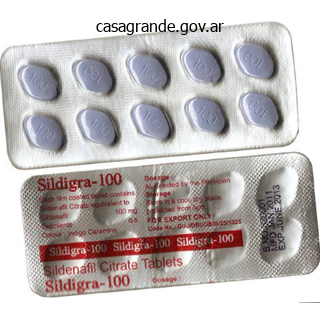
Sildigra dosages: 120 mg, 100 mg, 50 mg, 25 mg
Sildigra packs: 30 pills, 60 pills, 90 pills, 120 pills, 180 pills, 270 pills
Discount 50 mg sildigra amex
Cut part of the plaque reveals the luminal surface as a firm, white fibrous cap and a central core composed of yellow to yellow-white, delicate, porridgelike material and hence the name atheroma. Cellular space under the fibrous cap is comprised by a mix of macrophages, foam cells, lymphocytes and a few easy muscle cells which may comprise lipid. Deeper central delicate core consists of extracellular lipid material, cholesterol clefts, fibrin, necrotic debris and lipidladen foam cells. In older and extra advanced lesions, the collagen within the fibrous cap may be dense and hyalinised, smooth muscle cells could additionally be atrophic and foam cells are fewer. Various pathologic changes that happen in fully-developed atheromatous plaques are referred to as the sophisticated lesions. Calcification occurs extra commonly in advanced atheromatous plaques, particularly in the aorta and coronaries. The diseased intima cracks like an eggshell when the vessel is incised and opened. Microscopically, the calcium salts are deposited within the vicinity of necrotic area and in the delicate lipid pool deep within the thickened intima. The layers masking the gentle pultaceous materials of an atheroma could ulcerate as a outcome of haemodynamic forces or mechanical trauma. The ulcerated plaque and the areas of endothelial injury are susceptible sites for formation of superimposed thrombi. Intimal haemorrhage may happen in an atheromatous plaque both from the blood in the vascular lumen through an ulcerated plaque, or from rupture of thin-walled capillaries that vascularise the atheroma from adventitial vasa vasorum. Though atherosclerosis is primarily an intimal illness, superior lesions are associated with secondary adjustments in the media and adventitia. The modifications in media embrace atrophy and thinning of the media and fragmentation of inside elastic lamina. These changes cause weakening within the arterial wall leading to aneurysmal dilatation. Clinical Effects the medical effects of atherosclerosis depend upon the size and kind of arteries affected. Direct invasion of the artery by infectious brokers, especially micro organism and fungi, causes infectious arteritis. It is usually seen close to the lesions of peptic ulcers of the abdomen and duodenum, tuberculous and chronic abscesses in the lungs, persistent cutaneous ulcers, persistent meningitis, and in post-partum and post-menopausal uterine arteries. Microscopically, the obliteration of the lumen is as a end result of of concentric and symmetric proliferation of mobile fibrous tissue within the intima. Non-syphilitic Infective Arteritis Various types of invasions of the artery by bacteria, fungi, parasites or viruses, either instantly or by haematogenous route, trigger non-syphilitic infective arteritis. Syphilitic Arteritis Syphilitic or luetic vascular involvement happens in all stages of syphilis however is more prominent in the tertiary stage. Syphilitic involvement of the ascending aorta and the aortic arch is the most common manifestation of cardiovascular syphilis. Longitudinally opened vessels present intimal surface studded with pearlywhite thickenings, various from a few millimeters to a centimeter in diameter. These lesions are separated by wrinkled regular intima, giving it attribute tree-bark look. There is endarteritis and periarteritis of the vasa vasorum within the media and adventitia. Perivascular accumulation of plasma cells, lymphocytes and macrophages which will type miliary gummas which endure necrosis and are changed by scar tissue. Intimal thickenings include dense avascular collagen that may undergo hyalinisation and calcification. These are as follows: a) Aortic aneurysm might outcome from injury to the aortic wall (page 406). The aortic incompetence outcomes from spread of the syphilitic process to the aortic valve ring. The options distinguishing syphilitic aortitis from aortic atheroma are given in Table 15. Syphilitic involvement of small and mediumsized cerebral arteries occurs through the tertiary syphilis. Syphilitic Aortitis Ascending aorta, aortic arch; absent beneath diaphragm Pearly-white intimal lesions resembling tree-bark without fat within the core; ulceration and calcification typically not found Endarteritis and periarteritis of vasa vasorum, perivascular infiltrate of plasma cells and lymphocytes Thoracic aortic aneurysm, incompetence of the aortic valve, stenosis of coronary ostia Aortic Atheroma Progressive increase from the arch to stomach aorta, more typically at the bifurcation Yellowish-white intimal plaques with fat in the core; ulceration and calcification in plaques common Fibrous cap with deeper core containing foam cells, cholesterol clefts and gentle lipid Abdominal aortic aneurysm, aortic valve stenosis, stenosis of belly branches Feature 1. Most generally affected organs, in descending order of frequency of involvement, are the kidneys, heart, liver, gastrointestinal tract, muscle, pancreas, testes, nervous system and skin.
Generic sildigra 25mg amex
Normally, the nuclei of epithelial cells are oriented along the basement membrane which is termed as basal polarity. Early in malignancy, tumour cells lose their basal polarity in order that the nuclei are inclined to lie away from the basement membrane. The extent of cellular pleomorphism typically correlates with the diploma of anaplasia. Tumour cells are often bigger than normal however in some tumours they are often of normal dimension or smaller than regular. Nuclei are enlarged Diagrammatic representation of cytomorphologic features of neoplastic cells. Just like cellular pleomorphism, the nuclei too, present variation in size and form in malignant tumour cells. Characteristically, the nuclear chromatin of malignant cell is increased and coarsely clumped. This is as a result of of improve in the amount of nucleoprotein resulting in dark-staining nuclei, referred to as hyperchromatism. Nuclear shape may range, nuclear membrane may be irregular and nuclear chromatin is clumped along the nuclear membrane. Malignant cells frequently have a distinguished nucleolus or nucleoli within the nucleus reflecting elevated nucleoprotein synthesis. The parenchymal cells of poorlydifferentiated tumours typically present massive number of mitoses as compared with benign tumours and well-differentiated malignant tumours. Multinucleate tumour large cells or giant cells containing a single large and weird nucleus, possessing nuclear characters of the adjacent tumour cells, are one other important characteristic of anaplasia in malignant tumours. Structural anaplasia in tumours is accompanied with practical anaplasia as appreciated from the cytoplasmic constituents of the tumour cells. The functional abnormality in neoplasms may be quantitative, qualitative, or each. The new capillaries add to the vascular density of the tumour which has been used as a marker to assess the rate of development of tumours and hence grade the tumours. However, if the tumour outgrows its blood provide as happens in rapidly growing tumours or tumour angiogenesis fails, its core undergoes ischaemic necrosis. Inflammatory Reaction At instances, outstanding inflammatory response is present in and across the tumours. All tumour cells have abnormal genetic composition and on division they transmit the genetic abnormality to their progeny. Tumour Angiogenesis and Stroma the connective tissue alongwith its vascular community types the supportive framework on which the parenchymal tumour cells develop and obtain nourishment. In order to present nourishment to growing tumour, new blood vessels are shaped from Tumour stroma. B, Scirrhous carcinoma of breast having abundant collagenised (desmopastic) stroma. Generally, larger, more aggressive and rapidly-growing tumours are more likely to metastasise however there are quite a few exceptions. In general, carcinomas metastasise by lymphatic route whereas sarcomas favour haematogenous route. The involvement of lymph nodes by malignant cells could also be of two forms: i) Lymphatic permeation. The walls of lymphatics are readily invaded by cancer cells and will type a continuous development within the lymphatic channels known as lymphatic permeation. Alternatively, the malignant cells might detach to type tumour emboli in order to be carried alongside the lymph to the following draining lymph node. Generally, regional lymph nodes draining the tumour are invariably concerned producing regional nodal metastasis. Other instances, due to obstruction of the lymphatics by tumour cells, the lymph circulate is disturbed and tumour cells unfold in opposition to the circulate of lymph inflicting retrograde metastases at uncommon websites. This is due to cell-mediated immunologic response by the host in an attempt to destroy the tumour. Most benign tumours kind encapsulated or circumscribed masses that increase and push aside the surrounding regular tissues without really invading, infiltrating or metastasising.
Buy sildigra 50mg overnight delivery
Dose escalation For the first protocol in the dose escalation collection a reference dose of 10. The reference dose was defined as the dose to a 1 cm3 volume centered at the maximum of the thermal flux. The reference dose corresponded to the utmost dose, Dmax, when one subject was used. Encouraged by the results from the first 15 sufferers a model new protocol was began with a prescribed reference dose of 12. The reference dose was then outlined because the 1 cm3 centered on the maximum of the thermal flux exterior the tumor volume. In this new protocol a stratifying standards was established which separated the patients into two teams. If the goal could presumably be handled to a minimal dose of 17 Gy-Eq with one area the patient will be positioned on the one field group (protocol 4a). If the goal was too huge to be covered by the one subject then the affected person will be positioned on the double field group (protocol 4b). A vital enhance within the common brain dose was observed when two fields had been used as seen in determine 1. At this time new radiobiological research in animals have been commissioned to consider the boron concentration in different head and neck tissues in addition to to test the radiobiological effectiveness of this therapy on skin, mucosae, and glandular tissues. These studies revealed that there was a rise focus of boron in these head and neck tissues. As new information was uncovered the dose analysis for remedy planning was revised accordingly. All of them had subtherapeutic ranges of antiseizure treatment throughout and/or after the process. Another lesson discovered here was to keep the extent of antiseizure medicine therapeutic throughout and after the treatment. It is essential to increase the boron focus within the blood and subsequently in the tumor to take full advantage of this binary therapy. This protocol was designed to answer the question of attainable benefits of boron redistribution in tumor and potential changes in the tolerance of regular tissues after fractionation. Out of eleven sufferers accrued in these 4 protocols 7 have been placed in protocol 5, 2 in protocol 6 and one each in protocol 7 and 8. For the current protocols the target volume definition was changed from the gadolinium enhanced region plus a 2 cm shell round it, to the larger of the postoperative gadolinium enhanced area or the preoperative peritumoral edema and the two cm shell that encompasses it. This change mirrored our statement that every one recurrences occurred domestically and their progression followed the preoperative edema quantity sample and information from Kelly et. This included the volume of edema within the goal, the place the risk for recurrence is highest. It additionally made the volumes used to report our knowledge in preserving with the volumes used in the radiooncologic literature. It is too early to inform what the long term effects in sufferers accrued under the current protocols will be. Government retains a non-exclusive, royalty-free license to publish or reproduce the printed type of this contribution, or allow different to do so, for U. In Neuwelt (ed): Implication of the blood-brain barrier and its manipulation: Clinical elements, Volume 2 New York Plemum Press (1989), pp 77�106. Proceedings of the Sixth International Symposium on Neutron Capture Therapy for Cancer, Kobe, Japan October 31�November four, 1994, in Mishima Y (ed): Cancer Neutron Capture Therapy: New York, Plenum Press, 1996, pp 739�745. The dangers of central nervous system relapse and leukoencephalopathy in sufferers receiving marrow transplants for acute leukemia. A historical past of boron neutron capture remedy of brain tumor; Postulation of a brain radiation dose tolerance limit. Long term follow-up mind metastases handled by percutaneous stereotactic single high-dose irradiation. It is the first time that a medical software might be realised on a completely multi-national scale.
Purchase 100 mg sildigra mastercard
The suffix E or S are used for extranodal involvement and splenomegaly respectively (Table 14. Detailed bodily examination including websites of nodal involvement and splenomegaly. Chest radiograph to exclude mediastinal, pleural and lung parenchymal involvement. More invasive investigations embody lymphangiography of decrease extremities and staging laparotomy. Nodular sclerosis variety too has superb prognosis however those sufferers with larger mediastinal mass respond poorly to each chemotherapy and radiotherapy. Marrow examination shows malignant undifferentiated cells of precursor B or T cell origin as demonstrated by immunophenotyping. Cytochemical stains could additionally be employed as an adjunct to Romanowsky staining for determining the sort of leukaemia. TdT (terminal deoxynucleotidyl transferase) is expressed by the nuclei of both pre-B and pre-T levels of differentiation of lymphoid cells. Cases with lymphadenopathy at presentation present replacement of the lymph node by diffuse proliferation of well-differentiated, mature, small and uniform lymphocytes without any cytologic atypia or vital mitoses. These cells are of monoclonal B-cell origin having immunologic options of mantle zone B-cells. Prognosis typically correlates with the stage of disease as underneath: Stage A: characterised by lymphocytosis alone, or with limited lymphadenopathy, has an excellent prognosis (median survival greater than 10 years). Following options are seen: Lymph node biopsy: As the name suggests, follicular lymphoma is characterised by follicular or nodular pattern of progress. The nuclei of tumour cells could vary from predominantly small cleaved (or indented) to predominantly large cleaved selection. This selection is the diffuse counterpart of follicular giant cleaved cell lymphoma i. In common, diffuse large B-cell lymphomas are aggressive tumours and disseminate extensively. Tumour cells are intermediate in dimension, non-cleaved, and homogeneous in size and form. Mantle cell lymphoma arises from B-cells of mantle zone of regular lymphoid follicle. A, Peripheral blood shows presence of a leukaemic cells with furry cytoplasmic projections B, Trephine biopsy exhibits substitute of marrow spaces with irregular mononuclear cells. These cells are greatest recognised under part contrast microscopy but may be visible in routine blood smears. There is infiltration by IgM-secreting monoclonal lymphoplasmacytic cells into lymph nodes, spleen, bone marrow, and typically in the peripheral blood. The condition is commonly preceded by eczema or dermatitis for a number of years (premycotic stage). The disease could spread to viscera and to peripheral blood as a leukaemia characterised by S�zary cells having cerebriform nuclei termed as S�zary syndrome. Involvement of the skin occurs incessantly and produces an indolent cutaneous giant T/null cell lymphoma. These are more widespread in younger adults and infrequently have bone marrow involvement at presentation. The patients have profound constitutional signs (fever, weight loss, pores and skin rash), generalised lymphadenopathy and polyclonal hypergammaglobulinaemia. The situation is sort of aggressive and was earlier known as as deadly midline granuloma or angiocentric lymphoma. During the course of the disease, blood and bone marrow may be concerned producing leukaemic image. This staging system relies upon upon the number and location of nodal and extranodal websites involved, and presence or absence of constitutional (B) symptoms.
Purchase discount sildigra on-line
The subepithelium has fibromyxoid appearance and contains proliferating blood vessels a few of which are hyalinised. Sometimes, the subepithelial basement membrane is thickened, resembling amyloid materials. Generally, carcinoma of the supraglottic and subglottic regions tends to be more poorly-differentiated than the glottic tumour. Microscopically, the cyst is lined by stratified squamous or respiratory epithelium, overlaying subepithelial lymphoid tissue aggregates or follicles with germinal centres. Parathyroid cyst is a lateral cyst of the neck normally located deep to the sternocleidomastoid muscle on the angle of the mandible. Cystic hygroma is a lateral swelling at the root of the neck, usually situated behind the sternocleidomastoid muscle. Microscopically, cystic hygroma is a diffuse lymphangioma containing giant cavernous spaces lined by endothelium and containing lymph fluid (page 412). Primary Tumours A few essential examples of main tumours in the neck are carotid body tumour, torticollis and malignant lymphomas. Carotid body tumour arises within the carotid our bodies that are situated on the bifurcation of the common carotid arteries. Carotid our bodies 521 are usually part of the chemoreceptor system and the cells of this system are delicate to modifications within the pH and arterial oxygen rigidity and are additionally the storage website for catecholamines. Histologically similar tumours are found in other parasympathetic ganglia represented by the vagus and glomus jugulare (jugulotympanic our bodies, Chapter 27). Microscopically, well-differentiated tumour cells kind attribute organoid or alveolar sample, as is the case with all different neuroendocrine tumours. This is a deformity in which the pinnacle is bent to one aspect while the chin factors to the opposite facet. Secondary Tumours Cervical lymph nodes are frequent site for metastases of a lot of carcinomas. Characteristically, oral lichen planus appears as interlacing community of whitening or keratosis on the buccal mucosa however other oral tissues corresponding to gingival, tongue and palate can also be involved. Vesicular oral lesions appear invariably in all circumstances at some time in the course of pemphigus vulgaris. Vesicles or bullae seem on oral mucosa in addition to on conjunctiva in pemphigoid and are seen more often in older girls. Cleft higher lip (harelip) and cleft palate, alone or in combination, are the most typical developmental anomalies of the face. This is an asymptomatic condition occurring in kids and is characterised by symmetric, greywhite areas on the buccal mucosa. These are as beneath: i) Macroglossia is the enlargement of the tongue, usually due to lymphangioma or haemangioma, and generally due to amyloid tumour. It is the most typical manifestation of major an infection with herpes simplex virus. Recurrent assaults happen because of stress, emotional upsets and higher respiratory infections. The lesions are characterised by necrosis of the marginal gingiva and should lengthen on to oral mucosa, inflicting cellulitis of the tissue of the cheek. Cervicofacial actinomycosis is the most common type of the illness growing at the angle of the mandible (Chapter 6). Acute glossitis characterised by swollen papillae happens in eruptions of measles and scarlet fever. Oral lesions could happen in main, secondary, tertiary and congenital syphilis (Chapter 6). All forms of pigmented naevi in addition to malignant melanoma can happen in oral cavity. Fibrous growths (Fibroepithelial polyps, fibrous epulis, denture hyperplasia) Pyogenic granuloma Mucocele Ranula Dermoid cyst 2.
Cheap generic sildigra canada
Ischaemic necrosis may be as a result of embolism per se, but other components such as platelet activation, intravascular coagulation and hypoxia might contribute. Amniotic Fluid Embolism that is the most serious, unpredictable and unpreventible explanation for maternal mortality. During labour and within the quick postpartum interval, the contents of amniotic fluid could enter the uterine veins and attain right aspect of the guts leading to fatal problems. The medical syndrome of amniotic fluid embolism is characterised by the next features: Sudden respiratory misery and dyspnoea Deep cyanosis Cardiovascular shock Convulsions Coma nexpected dying the cause of death will not be obvious but can happen on account of the next mechanisms: i) Mechanical blockage of the pulmonary circulation in in depth embolism. Atheroembolism Atheromatous plaques, particularly from aorta, may get eroded to form atherosclerotic emboli which are then lodged in medium-sized and small arteries. Pathologic adjustments and their effects in atheroembolism are as underneath: i) Ischaemia, atrophy and necrosis of tissue distal to the occluded vessel. Notable examples are clear cell carcinoma of kidney, carcinoma of the lung, malignant melanoma and so on (Chapter 8). These are: i) Fragments of tissue ii) Placental fragments iii) Red cell aggregates (sludging) iv) Bacteria v) Parasites vi) Barium emboli following enema vii) Foreign our bodies. The cessation of blood provide may be full (complete ischaemia) or partial (partial ischaemia). Hypoxia as a outcome of deprivation of oxygen to tissues; that is crucial and common cause. Inadequate clearance of metabolites which outcomes in accumulation of metabolic waste-products within the affected tissue; that is related in some conditions similar to muscleache after ischaemia from heavy train. These causes are discussed below with regard to different levels of blood vessels: 1. Inadequate cardiac output resulting from coronary heart block, ventricular arrest and fibrillation from various causes could trigger hypoxic injury to the mind. The commonest and most essential causes of ischaemia are because of obstruction in arterial blood provide. These are as beneath: i) Luminal occlusion of artery: a) Thrombosis b) Embolism ii) Causes in the arterial walls: a) Vasospasm. Blockage of venous drainage might lead to engorgement and obstruction to arterial blood supply resulting in ischaemia. The examples embody the next: i) Luminal occlusion of vein: a) Thrombosis of mesenteric veins b) Cavernous sinus thrombosis ii) Causes within the vessel wall of vein: a) Varicose veins of the legs iii) Outside stress on vein: a) Strangulated hernia b) Intussusception c) Volvulus four. The causes are as underneath: i) Luminal occlusion in microvasculature: a) By red cells. The extent of damage produced by ischaemia because of occlusion of arterial or venous blood vessels depends upon numerous factors. The extent of injury by ischaemia relies upon upon the anatomic sample of arterial blood supply of the organ or tissue affected. There are 4 totally different patterns of arterial blood supply: i) Single arterial provide with out anastomosis. Arterial provide to some organs has rich interarterial anastomoses in order that blockage of 1 vessel can re-establish blood provide bypassing the blocked arterial branch, and therefore the infarction is less widespread in such circumstances. Interarterial anastomoses in the three major trunks of the a hundred twenty five coronary arterial system. Blood provide to some organs and tissues is such that the vitality of the tissue is maintained by alternative blood provide in case of occlusion of one. The impact of occlusion of one set of vessels is modified if an organ has dual blood supply. For instance: Lungs are perfused by bronchial circulation as properly as by pulmonary arterial branches. The basic status of a person as regards cardiovascular function is a crucial determinant to assess the impact of ischaemia. The mesenchymal tissues are fairly immune to the impact of ischaemia as compared to parenchymal cells of the organs. Complete vascular obstruction leads to more severe ischaemic injury than the partial occlusion. No results on the tissues, if the collateral channels develop adequately in order that the effect of ischaemia fails to happen. Some basic morphological features of infarcts are frequent to infarcts of all organ websites.
Diseases
- Granulomatous rosacea
- Congenital adrenal hyperplasia, lipoid
- Mucormycosis
- Granulomas, congenital cerebral
- Retinis pigmentosa deafness hypogenitalism
- Urticaria
Order sildigra online now
The hypoperfusion of the alimentary tract in conditions such as shock and cardiac failure might lead to mucosal and mural infarction called haemorrhagic gastroenteropathy (Chapter 20). This kind of non-occlusive ischaemic damage of bowel should be distinguished from full-fledged infarction by which case the deeper layers of intestine (muscularis and serosa) are also broken. Microscopically, relying upon the time lapse between damage and cell demise, ischaemic shrinkage, hydropic change, focal necrosis, or fatty change could also be seen. Other organs corresponding to lymph nodes, spleen and pancreas may show foci of necrosis in shock. In addition, the patients who survive acute part of shock succumb to overwhelming an infection as a end result of altered immune status and host protection mechanism. Clinical Features and Complications the classical features of decompensated shock are characterised by despair of four vital processes: Very low blood pressure Subnormal temperature Feeble and irregular pulse Shallow and sighing respiration In addition, the sufferers in shock have pale face, sunken eyes, weak spot, cold and clammy pores and skin. Life-threatening problems in shock are as a end result of hypoxic cell harm resulting in immuno-inflammatory responses and activation of various cascades (clotting, complement, kinin). Haemostatic plugs are the blood clots formed in healthy individuals on the web site of bleeding. In other phrases, haemostatic plug at the cut finish of a blood vessel may be considered the best form of thrombosis. Haemostatic plugs are helpful as they cease the escape of blood and plasma, whereas thrombi growing within the unruptured cardiovascular system could also be life-threatening by causing one of many following dangerous results: 1. Thrombi might decrease or cease the blood provide to part of an organ or tissue and trigger ischaemia which may subsequently end in infarction. The thrombus or its half could get dislodged and be carried alongside in the bloodstream as embolus to lodge in a distant vessel. Human beings possess inbuilt system by which the blood remains in fluid state normally and guards against the hazards of thrombosis and haemorrhage. However, damage to the blood vessel initiates haemostatic repair mechanism or thrombogenesis. To this are added the processes that follow these primary occasions: activation of platelets and of clotting system. The integrity of blood vessel wall is important for maintaining normal blood move. An intact endothelium has the next features: i) It protects the flowing blood from the thrombogenic influence of subendothelium. B, Endothelial harm exposes subendothelium, initiating adherence of platelets and activation of coagulation system. D, Activated coagulation system varieties fibrin strands by which are entangled some leucocytes and purple cells and a decent meshwork is fashioned referred to as thrombus. A, Normal non-activated platelet, having open canalicular system and the cytoplasmic organelles dispersed within the cell. B, Early adhesion part, showing dilatation of the canalicular system with formation of pseudopods and the organelles current in the centre of the cell. Endothelial damage is of main significance within the formation of arterial thrombi and thrombi of the center, particularly of the left ventricle. A variety of components and conditions may trigger vascular injury and predispose to the formation of thrombi. These are as under: i) Endocardial harm in myocardial infarction, myocarditis, cardiac surgical procedure, prosthetic valves. Following endothelial cell harm, platelets come to play a central position in normal haemostasis as well as in thrombosis. Normal non-activated platelets have open canalicular system with cytoplasmic organelles (granules, mitochondria, endoplasmic reticulum) dispersed all through the cytoplasm. The activated platelets then endure launch reaction by which the platelet granules are launched to the outside. Two primary kinds of platelet granules are launched: a) Alpha granules containing fibrinogen, fibronectin, plateletderived progress issue, platelet factor 4 (an antiheparin) and cationic proteins.
50mg sildigra fast delivery
The definitive center cerebral arteries comply with the indented surfaces of the insulae, first dipping into and then out of the sylvian fissures to ramify over the lateral surfaces of the frontal, parietal, and temporal lobes. After the sylvian fissures form (35-7), the subsequent groups of floor indentations to appear are the calcarine and parietooccipital sulci (35-8), followed by the central sulci (359). Gyral development occurs most quickly across the sensorimotor and visible pathways. Errors in Neuronal Migration and Cortical Organization the primary result of errors at these phases are malformations of cortical development. Examples include microcephaly, megalencephaly, heterotopias, cortical dysplasias, and lissencephaly. Operculization, Sulcation, and Gyration Lobulation and Operculization the cerebral hemispheres first appear as outpouchings of the embryonic telencephalon. The fetal cerebral vasculature covers the mind surface in a basket-like network of thin-walled, undifferentiated vessels. The cerebral hemispheres increase, first overlaying the diencephalon after which the midbrain and hindbrain. As the hemispheres elongate and rotate, they assume a "C" form with the caudal ends turning ventrally to kind the temporal lobes. Anomalies in Sulcation and Gyration Developmental errors in operculization, sulcation, and gyration are relatively unusual. Myelination Myelination happens in an orderly, predictable manner and could be detected as early as 20 fetal weeks. Normal mind myelination patterns in addition to abnormalities in myelin formation and maintenance are discussed in greater element in Chapter 31. In general, myelination proceeds from inferior to superior, from back to front, and from central to peripheral. Embryologically, the cerebellum is an extension of the midline and thus part of the dorsal pons. The rhombencephalic lips fuse, forming the cerebellar commissures in the roof of the fourth ventricle. A ridge of creating choroid plexus divides the rising fourth ventricle into anterior and posterior membranous areas. Normally, the anterior membrane is integrated into the growing choroid plexus, whereas the posterior membranous space persists and ultimately cavitates, forming the midline foramen of Magendie. Midbrain and Hindbrain Development We now summarize the main embryologic events involved in forming the midbrain and hindbrain. Midbrain-Hindbrain Anomalies A variety of completely different classifications of midbrain and hindbrain malformations have been proposed. Such a system makes consummate sense and definitely aids understanding the pathogenesis of these fascinating anomalies. However, the more conventional morphologic-based strategy by which malformations are grouped according to imaging findings is the simplest for radiologists to comply with. The interested reader is referred to the publications cited at the finish of Chapter 36. Major Embryologic Events the mesencephalon gives rise to the brainstem, and the rhombencephalon elaborates into the medulla, pons, and cerebellum. The mesencephalon is divided into ventral (tegmentum) and dorsal (tectum) areas. Likewise, the metencephalon is divided into ventral (pons) and dorsal (cerebellum) regions. The pons is formed by a proliferation of cells and fiber tracts along the ventral metencephalon. Thin cortex and shallow, incompletely formed sulci are normal for such a untimely infant. The medulla has myelinated and elevated in signal depth, as have the flocculi and dentate nuclei. Although parenchymal calcifications, ventricular size/configuration, and main abnormalities may be recognized, subtle abnormalities corresponding to cortical dysplasia are troublesome to detect and straightforward to overlook.
Sildigra 25 mg lowest price
Fibronectin varieties part of the extracellular matrix between cells that "glues" them together. Decreased manufacturing of tissue plasminogen activator from intact endothelial cells might occur in anoxia of the endothelial cells in veins with sluggish circulation. Stasis of blood circulate is essential in thrombosis throughout the low-pressure venous circulation. Thromboxane A2 is released when platelets are activated through the strategy of platelet adhesion. They are vitamin K dependent and due to this fact can also be affected by warfarin remedy or by parenchymal liver illness. The issue V Leiden mutation leads to problem inactivating factor V by the action of protein C, thus causing thrombosis, not bleeding. Scurvy from vitamin C deficiency affects vascular integrity, not coagulation elements. The issue V (Leiden) mutation affects 2% to 15% of the population, and greater than half of all people with a historical past of recurrent deep venous thrombosis have such a defect. Hyperhomocysteinemia is a much less widespread reason for inherited threat of thrombosis than is issue V mutation. It is also a threat factor for atherosclerosis that predisposes to arterial thrombosis. Although some cancers elaborate components that promote thrombosis, this affected person is unlikely to have most cancers at such a younger age; a 10-year historical past of thrombosis is unlikely to happen in a patient with most cancers. Oral contraceptive usage contributes to danger for thrombosis, but mainly in older women, significantly previous age 40 years. Aspirin prevents formation of recent thrombi by inhibiting platelet aggregation and works finest as preventive therapy. The patient has antibodies that react with cardiolipin, a phospholipid antigen used for the serologic prognosis of syphilis. Patients with antiphospholipid syndrome have recurrent arterial and venous thrombosis and repeated miscarriages. Hypercholesterolemia promotes atherosclerosis over many years, and the danger of arterial thrombosis increases. Von Willebrand illness impacts platelet adhesion and leads to a bleeding tendency, to not thrombosis. Diabetes mellitus accelerates and worsens atherosclerosis, together with peripheral muscular arteries. The embolus is carried distally and should occlude the popliteal artery, which is an end artery for the lower leg. A vegetation is a localized thrombus formation on cardiac endothelium, sometimes a valve. Calcium is a cofactor within the coagulation pathway, but an increase in calcium has minimal effect on the coagulation process. Turbulent blood flow might promote thrombosis, but this threat factor is more frequent in fast-flowing arterial circulation. Fat embolism can occur with fractures, but pulmonary issues usually seem 1 to 3 days after the traumatic occasion. Vessels with thrombi usually stay intact; if a hematoma had developed as a consequence of the trauma from the fall, it will be organizing and decreasing in size after 2 weeks. Such a peripheral arterial occlusion was inadequate to produce infarction, as evidenced by the dearth of enzyme elevation. After a thrombus has fashioned, it might become organized with ingrowth of capillaries, fibroblast proliferation, and macrophage infiltration, which finally clears part or a lot of the clot; there could be formation of one or more new lumens (recanalization). Air emboli on the arterial facet could cause ischemia via occlusion even when very small, whereas on the venous side, more than 100 mL of air trapped within the heart might scale back cardiac output. When gases that turned dissolved in tissues at high stress bubble out at decompression with lower pressure in blood and tissues, then air emboli form.
Buy discount sildigra 100 mg on line
An acute lung abscess is initially surrounded by acute pneumonia and has poorlydefined ragged wall. Histologically, the attribute characteristic is the destruction of lung parenchyma with suppurative exudate in the lung cavity. The scientific manifestations are fever, malaise, loss of weight, cough, purulent expectoration and haemoptysis in half the cases. These infections in healthy people are rarely serious but in immunosuppressed people might prove fatal. Aspergillosis is the most common fungal an infection of the lung caused by Aspergillus fumigatus that grows best in cool, wet climate. Candidiasis or moniliasis brought on by Candida albicans is a normal commensal in oral cavity, gut and vagina but attains pathologic type in immunocompromised host. It is attributable to oval organism, Histoplasma capsulatum, by inhalation of contaminated dust or chook droppings. It is brought on by Cryptococcus neoformans which is round yeast having a halo around it as a result of shrinkage in tissue sections. Pulmonary lesions attributable to Mycobacterium tuberculosis and other mycobacteria have already been discussed along with common aspects of tuberculosis and different granulomatous inflammations in Chapter 6. Bronchiectasis Chronic bronchitis and emphysema are quite frequent and often happen collectively. The two most necessary etiologic components responsible for majority of circumstances of continual bronchitis are: cigarette smoking and atmospheric pollution. The mostly identified factor implicated in causation of continual bronchitis and in emphysema is heavy smoking. Prolonged cigarette smoking seems to act on the lungs in a quantity of methods: i) It impairs ciliary movement. The Respiratory System 478 iv) It causes appreciable obstruction of small airways. The incidence of chronic bronchitis is larger in industrialised urban areas the place air is polluted. Workers engaged in sure occupations such as in cotton mills (byssinosis), plastic factories etc. Cigarette smoke, however, predisposes to infection liable for acute exacerbation in chronic bronchitis. There appears to be a poorly-defined familial tendency and genetic predisposition to develop disabling persistent bronchitis. Lumina of the bronchi and bronchioles could contain mucus plugs and purulent exudate. There is appreciable overlap of medical features of chronic bronchitis and pulmonary emphysema (discussed below) as very often the two coexist. As mentioned to begin with of this chapter, a lobule is composed of about 5 acini distal to a terminal bronchiole and that an acinus consists of three to 5 generations of respiratory bronchioles and a variable variety of alveolar ducts and alveolar sacs (page 461). The association of the two situations is principally linked to the frequent etiologic factors- most significantly tobacco smoke and air pollutants. Other much less significant contributory factors are occupational exposure, infection and somewhat poorly-understood familial and genetic influences. Clinically significant deficiency is also related to homozygous Pi null null and heterozygous Pi nullZ. The mechanism of alveolar wall destruction in emphysema by elastolytic motion relies on the imbalance between proteases (chiefly elastase) and anti-proteases (chiefly anti-elastase): By decreased anti-elastase activity i. There are sufficient evidences to suggest that smoking promotes emphysema by both lowering the quantity of antielastase in addition to by growing the elastolytic protease within the lungs. The Respiratory System 480 Grossly, the lungs are voluminous, pale with little blood. The bullae are air-filled cyst-like or bubble-like constructions, bigger than 1 cm in diameter. They are formed by the rupture of adjoining air areas while blebs are the outcomes of rupture of alveoli directly into the subpleural interstitial tissue and are the frequent explanation for spontaneous pneumothorax. Emphysema may be identified with certainty only by gross and histologic examination of sections of entire lung.
Real Experiences: Customer Reviews on Sildigra
Goran, 53 years: Carbon monoxide binds far more tightly to hemoglobin than does oxygen, leading to hypoxia. Which of the following problems of this disease is more than likely to occur within 1 hour of those events Myocardial rupture Pericarditis Valvular insufficiency Ventricular fibrillation Thromboembolism 32 A 59-year-old man has experienced persistent fatigue for the past 18 months.
Rufus, 63 years: Blood glucose levels are tightly regulated and are normally maintained within a narrow physiologic vary. These modifications embrace segmental inflammation and fibrinoid change in the walls of capillaries.
8 of 10 - Review by N. Iomar
Votes: 158 votes
Total customer reviews: 158
References
- Trinh, T.A. and T.T.A. Vo, Evaluating the powerful prediction of integrated behavioral model for risky road behaviors. Procedia Eng., 2016.
- Timms D, Moss JP. An histological investigation into the effects of rapid maxillary expansion on the teeth and their supporting tissues. Trans Eur Orthod Soc 1971:263-271.
- Fukui M. Members of the Research Committee on Spontaneous Occlusion of the Circle of Willis (Moyamoya Disease) of the Ministry of Health and Welfare, Japan. Guidelines for the diagnosis and treatment of spontaneous occlusion of the circe of Willis ('moyamoya' disease). Clin Neurol Neurosurg 1997;99(Suppl. 2):S238-40.
- Elesber A, Lerman A, Bybee KA, et al. Myocardial perfusion in apical ballooning syndrome correlate of myocardial injury. Am Heart J. 2006;152:469.
- Miller WL: Fetal endocrine therapy for congenital adrenal hyperplasia should not be done, Best Pract Res Clin Endocrinol Metab 29(3):469n483, 2015.


