Gretchen M. Brophy, PharmD, BCPS, FCCP, FCCM, FNCS
- Professor of Pharmacotherapy & Outcomes Science and Neurosurgery, School of Pharmacy, Virginia Commonwealth University, Richmond, Virginia

https://www.usfq.edu.ec/eventos/neurocritico/Documents/gretchen_brophy.html
Sarafem dosages: 20 mg, 10 mg
Sarafem packs: 30 pills, 60 pills, 90 pills, 120 pills, 180 pills, 270 pills, 360 pills
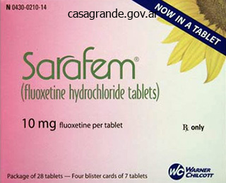
Cheap 10mg sarafem otc
A relative contraindication for this anatomic repair is generalized ligamentous laxity as may be encountered in Ehlers-Danlos syndrome. If an osteochondral lesion is present, the ligamentous reconstruction must be accomplished along side arthroscopic or open treatment of the osteochondral defect. This method facilitates entry to the peroneal tendons should there be related peroneal tendon pathology. The affected person is positioned as described, a thigh tourniquet is placed, and a normal orthopaedic prep and drape is carried out. With the bump placed proximal to the ankle, a dissection is carried out to isolate the inferior extensor retinaculum. The joint capsule is then incised in line with the skin incision and simply distal to the vanguard of the fibula. This inspection, together with the preoperative evaluation, is used to determine whether or not or not a restore of this ligament is needed. A subperiosteal dissection is carried out on the anterior and lateral side of the fibula, elevating a flap 3 to 6 mm broad. Using curettes and rongeurs, a trough is made within the anterior and lateral side of the fibula at its vanguard, about 3 mm deep and 3 mm broad. If additional shortening is needed, the capsule may be trimmed from the distal cut edge. A second reinforcing layer of restore is created by suturing the inferior extensor retinaculum to the periosteal flap with absorbable 2-0 figure eight sutures. The skin is closed in layers with 3-0 absorbable suture within the subcutaneous suture and staples or subcuticular suture used within the pores and skin. Modified Brostrom Anatomic Lateral Ankle Ligament Repair with Suture Anchor(s) (Courtesy of Mark E. Lift the limb by the anchors; if the anchors are going to fail, we would like them to accomplish that now so the issue can be rectified. Mobilize the inferior extensor retinaculum to be used to augment the repair (Gould modification of the Brostrom procedure). Stability of suture anchors tested by lifting limb from the operating room table by the anchor sutures. Reduce the talus throughout the ankle mortise before reattaching the ligaments and capsule. The ankle is held in dorsiflexion, with a posterior pressure maintaining the talus inside the ankle mortise. Although coated, a bump has been positioned underneath the distal tibia to permit the heel to translate posteriorly without interfering with the working table. Recheck the anterior drawer take a look at to decide if the first sutures are securely sustaining ankle stability. Final verify of anterior drawer and talar tilt tests to be sure repair is satisfactory. Lateral ankle instability in a patient with pre-exisiting longitudinal split tear of the peroneus brevis. Prepare the lateral ankle ligament advanced as is completed for the isolated modified Brostrom process. Transect the anterior 50% of the tendon proximally and cross this half of the peroneus brevis tendon beneath the intact superficial peroneal retinaculum. Pass the anterior slip of the peroneus brevis by way of the tunnel from distal to proximal. After passing by way of the fibular tunnel, the anterior slip of the peroneus brevis may be folded distally over the fibula to increase the repair. A bolster under the ipsilateral hip ensures that the leg is maintained in the optimal position, thereby maintaining adequate publicity to the lateral ankle. A bolster under the operated ankle can additionally be helpful and improves access to the lateral ankle.
Generic 20 mg sarafem with mastercard
The extremity is ready and draped, including the iliac crest if structural autograft is desired. Approach Traditionally, an extensile lateral strategy to the ankle and subtalar joints is used, though a posterior strategy has also been described. Tibiotalocalcaneal fusion is indicated in sufferers with arthritis in both the tibiotalar and subtalar joints. The aim of surgical intervention is to obtain a secure, plantigrade, and pain-free foot and ankle. These embrace (1) to remove medial bony prominences and debris and (2) to assist in resection of medial bone when advanced varus deformity precludes discount of the foot to impartial. Enter the ankle joint sharply and totally expose it by releasing the lateral ligaments and anterior and posterior capsule. After eradicating the cartilage, put together the joint floor with flexible chisels or a small, low-speed burr. Burr holes ought to be simply through the subchondral bone and separated by about three mm on all sides to avoid weakening or fracture of the cortex. Curette the remaining cartilage off the joint surface and put together the subchondral bone with versatile chisels or a burr as described above. In this case, the calcaneal articular processes might want to be removed with an osteotome to create a flat surface that may lie flush with the tibial plafond. The use of each an adolescent blade plate and a humeral blade plate has been described. Ensure that the hindfoot is positioned in neutral to 5 degrees of valgus and the ankle is in impartial dorsiflexion and plantarflexion. The ankle and subtalar joints must be held rigidly throughout insertion of the blade plate. The guidewire must be inserted such that 5 to 10 mm of calcaneal bone will stay plantar to the blade. The lateral calcaneal cortex could then be additional ready for blade insertion by predrilling with a four. Contour the plate to the lateral facet of the tibia and fill the screw holes sequentially. Place the screw under fluoroscopic guidance from the calcaneal tuberosity into the anterior tibial cortex at roughly a 60-degree angle. Further steps that may help in the prevention of a postoperative hematoma include releasing the tourniquet and assessing hemostasis before closure, the use of drains, and the utilization of a compression dressing. Resecting a groove for the plate in essentially the most distal portion of the tibia utilizing a chisel or burr can also be helpful. Failure to contour the blade plate when necessary can sometimes push the hindfoot into varus. In instances of talectomy, the foot have to be aligned with the leg, as alignment of the distal tibia to the posterior calcaneal aspect can outcome in a dorsiflexion malunion. After 10 to 14 days, patients return to the office for evaluation of the wound and suture removing. Patients stay non�weight-bearing in a short-leg solid for 6 to 12 weeks, based mostly on radiographic healing. Thereafter, sufferers are transitioned to a short-leg walking forged or boot and progressive weight bearing is begun. In studies examining using blade plate fixation solely, the reported fusion rates have ranged from 90% to one hundred pc. In this case, the talus was "replaced" with a carefully contoured femoral head allograft. In patients present process tibiotalocalcaneal fusion (regardless of fixation technique) the nonunion price ranges from 0% to 40%. Peripheral neuroma of both the sural or superficial peroneal nerves may be minimized by cautious incision placement and delicate retraction and soft tissue handling. Intramedullary rod fixation in contrast with blade-plate-and-screw fixation for tibiocalcaneal arthrodesis: a biomechanical investigation.
10mg sarafem
It is essential to inspect all pin websites every day to assess for indicators of infection or loosening, together with localized redness, ache and tenderness, heat, swelling (firm or fluctuant), and drainage from the pin or wire that may vary in shade and odor. When early signs of pin web site infection are noted, pin care is increased to twice every day, the pin site is wrapped with a gauze roll dressing, ankle vary of motion is discontinued, and weight bearing and physical therapy are restricted. The pin site infection normally begins to resolve within 24 hours of beginning oral antibiotic remedy. Recalcitrant pin web site an infection is treated with intravenous antibiotic remedy with or with out pin removing. Physical therapy is began in the hospital and continued until after frame removal. Non-impact actions, including swimming and pool remedy if available, are inspired with the fixator in place. After adequate therapeutic of an osteotomy, nonunion, Achilles lengthening, or ligament reconstruction, ankle range of motion may be initiated with attention to optimize dorsiflexion. Ankle range of movement is initially done for half-hour, three to five occasions per day, and then progressed as tolerated by the affected person and pin sites. With related correction of equinus or varus deformity, quick distraction is prevented, and gradual distraction (0. Frame elimination: the body usually is eliminated 12 weeks after application beneath basic anesthesia as outpatient surgery. This could also be delayed till healing of a simultaneous osteotomy, nonunion correction, or malunion correction. After frame removal, gentle dressings are applied and adjusted as wanted for bleeding. Crutches or other assistive devices are used for the primary a quantity of weeks after body removing for comfort, but weight bearing as tolerated is allowed. Showers are resumed after the pin sites stop draining, normally within 3 days after frame removing. Care after body removal: During the preliminary 6 to 8 weeks after frame removal, the affected person steadily resumes common footwear and full weight bearing without assistive gadgets. Maintaining ankle range of motion, especially dorsiflexion, could also be facilitated with non-impact activities similar to swimming, bicycling, and physical remedy. A detachable fracture walker boot, compression stocking, or light ankle brace could minimize discomfort and swelling. In 11 sufferers with ankle arthritis associated with distal tibial deformity, remedy consisted of ankle joint distraction with the Ilizarov system, osteotomy, and range-of-motion workout routines for three months. It was concluded that deformity correction may augment the efficacy of distraction and that dorsiflexion could additionally be an necessary issue in the success of the ankle distraction procedure. A unfastened wire can often be retensioned within the office, whereas a free half-pin can only be removed in the workplace. Frame modification is sometimes needed if a quantity of half-pins become loose in the tibia early in the course of the distraction period. Most broken hardware such because the common hinges or threaded rods can be repaired within the office. The most significant problems of ankle distraction arthroplasty are failure to relieve ache and a lack of ankle movement. As a rule, swelling and stiffness do occur after ankle distraction as a end result of the underlying arthritis. A period of elevated pain and incapacity after ankle distraction could happen for 2 to 4 months after frame removal, often persisting for up to 6 to 12 months. Physical therapy and non-impact actions are emphasized throughout this time, together with swimming and bicycling. Patients should be counseled to wait a minimum of 12 months earlier than judging the success or failure of ankle distraction arthroplasty. Direct neurovascular damage ensuing from pin placement could happen despite operative warning because of posttraumatic distortion of the anatomy and scarring.
Purchase sarafem 10mg line
While the screws could appear distinguished, two-dimensional fluoroscopy is deceiving since the screws are countersunk beneath the articular surface of the graft and the talar dome is curved. The hardware may seem slightly proud fluoroscopically regardless of being countersunk. Moreover, the articular cartilage is somewhat thick compared to such a low-profile screw head. Confirm the reduction by way of the anteromedial arthrotomy and posteriorly behind the posterior tibial tendon. While not important for therapeutic, we favor placing an antiglide plate over the proximal facet of the osteotomy. There might be a slight hole on the medial malleolar osteotomy site regardless of anatomic reduction of the medial malleolus. Closure Posterior tibial tendon sheath and flexor retinaculum Anterior arthrotomy Subcutaneous layer Skin to a tensionless closure We routinely use a drain. Before continuing to the operating room, confirm that the allograft talus is the one intended for this affected person, is out there, and has not expired. Unlike ankle arthrodesis and complete ankle arthroplasty, should defend ankle cartilage. Since the whole medial one third to one half of the talar dome will be restructured, a medial malleolar osteotomy is typically not essential. If the talus seems appropriate for an allograft talus, ask to have the donor talus opened and soaking in a warm saline-soaked sponge on the back desk. We use a thin oscillating saw for this reduce, also with chilly saline irrigation to cool the blade in an try to avoid warmth necrosis to the bone. Fluoroscopic analysis sometimes affords a helpful appreciation of the recipient site. Harvesting Graft from the Donor Talus Secure the allograft that has been placed on the back desk with a bone-holding forceps. Further extraction of diseased cartilage until healthy-appearing cancellous surface is apparent. Attempt to match the recipient site dimensions exactly, considering the thickness of the noticed blade. Because the talus is contained within the ankle mortise, in our experience posterior screw fixation is pointless. We have never had an ideal match on the first try at seating the graft within the recipient web site. In our hands this requires a slight deepening of the recipient site and a slight thinning of the graft. Making the corresponding sagittal and axial talar cuts congruently is an important step in achieving an optimal fit of the graft. The human talus is quite variable and regardless of the match, some inconsistencies will be current. While the scientific look may counsel a nearperfect match, we routinely see slight incongruencies in the sagittal and axial preparations and what seems to be a slight mismatch to the native subchondral bone. These are positioned anteriorly and countersunk under the articular surface, usually anterior to the tibial plafond with the ankle in impartial place. Axial Realignment Based on the preoperative plan and intraoperative reassessment, contemplate correction of axial malalignment. Through the identical incision, perform supramalleolar osteotomy for varus malalignment. If hinge is weak, keep proper contact; management rotation of two fragments; consider using two plates in two planes for fixation. After additional "touch-ups" to the graft and recipient web site, optimal graft place. Stabilizing graft to native talus (blunt retractor superiorly and bone reduction clamp for coronal compression). Osteotomy being rigorously opened with an osteotome while preserving the lateral cortical hinge. Reapproximate the extensor retinaculum while defending the deep neurovascular bundle, extensor tendons, and the superficial peroneal nerve. Be certain the tissue financial institution leaves the cartilage on the talus (we have had tali delivered from tissue banks that routinely take away the cartilage from the allograft talus! Take care to orient the donor talus properly, as it ought to rest in the ankle mortise (compare to the native talus).
Order discount sarafem
The knee flexion check is carried out with the affected person susceptible and ankles clear of the table. Plain lateral radiographs may reveal an irregular configuration of the fat-filled triangular space anterior to the Achilles tendon and between the posterior side of the tibia and superior facet of the calcaneus (this house is named the triangle of Kager). However, sudden unexpected dorsiflexion of the ankle or violent dorsiflexion of a plantarflexed foot can also lead to ruptures. They generally complain of a limp and difficulties with actions of every day residing, significantly ascending stairs. This hole could also be absent in chronic ruptures, as the gap is often bridged by scar tissue. Active plantarflexion of the foot is often preserved because of the motion of tibialis posterior, the peroneal tendons, and the long toe flexors. If the gap in most plantarflexion is 5 to 9 cm, peroneus brevis switch can be utilized. Approach the normal midline longitudinal method over the Achilles tendon has been related to wound therapeutic issues and a risk of sural nerve harm when extended proximally. We employ a 10- to 12-cm curvilinear approach medial to the medial border of the tendon with sharp dissection via the subcutaneous fats to the paratenon. Positioning Under basic anesthesia, the affected person is positioned prone with the ankles clear of the operating desk. The ends of the Achilles tendon are freshened by sharp dissection, producing a defect between the freshened ends. Through the base of the wound, the deep fascia overlying the deep flexor compartment and the compartment containing the peronei muscular tissues may be seen. The internervous aircraft lies between the peroneus brevis (supplied by the superficial peroneal nerve) and the flexor hallucis longus (supplied by the tibial nerve). The tendons of the peroneus longus and brevis could be distinguished from one another at this level by the truth that though both are tendinous within the distal third of the lower leg, the peroneus brevis is muscular more distally than the peroneus longus. The deep fascia overlying the peroneal tendons is incised and the peroneal tendons are mobilized. Identify the peroneus brevis tendon, place a stay suture within the distal end of the peroneus brevis tendon, and detach the tendon from its insertion and mobilize it proximally. In this fashion, the tendon of the peroneus brevis retains its blood provide from the intermuscular septum. Incision remodeled insertion of peroneus brevis on the bottom of the fifth metatarsal. First move it from lateral to medial via the distal stump via coronal incisions medially and laterally within the Achilles tendon. Suture the sides of the coronal incisions in the Achilles tendon to the peroneus brevis tendon to stop progression of the incision that may lead to the peroneal tendon chopping out by way of the Achilles tendon. Pass the tendon via the proximal stump from medial to lateral, with the foot maximally plantarflexed. In most circumstances of neglected ruptures of the Achilles tendon, the paratenon is both not current or not viable. The paratenon, if not disrupted, is incised longitudinally within the midline for the length of the skin incision. If the remaining hole within the Achilles tendon is bigger than 9 cm, we proceed to harvest the gracilis tendon. Make a vertical 2- to 3-cm longitudinal incision on the medial facet of the tibial tuberosity, centered over the distal insertion of the pes anserinus. A venous plexus is usually encountered on the distal end of the wound, and care should be taken to diathermy this. Carry out dissection deep to the fats each medially and superiorly with a small swab on an artery forceps to expose the sartorius fascia. Insert a curved retractor and make a curved incision, 1 cm long, alongside the superior margin of the pes anserinus into the sartorius fascia, taking care to keep away from the saphenous nerve.
Syndromes
- Small eyes
- Bladder stones with prostate enlargement
- Sickle Cell Disease Association of America - www.sicklecelldisease.org
- Infection
- Paleness or dry skin
- Is there anything you do to reduce or prevent accidents?
- Testicular biopsy
- Headache
Purchase genuine sarafem line
For instance, when utilizing a nerve stimulator to administer regional anesthesia, an anesthesiologist could wish to document the length and kind of needle used, the nerve stimulator kind and settings, the variety of attempts, the strength and site of muscle contractions, whether paresthesia occurred, and the way the paresthesia was managed. Assume that a affected person had no urine output over a certain period in a beforehand functioning bladder catheter. The anesthesiologist treated the decreased urine output by the intravenous administration of crystalloid. The document would ideally point out "zero" underneath urine output and indicate a bolus of fluid beneath fluid administration. Assume, then, that half-hour after the crystalloid administration, the affected person still had no urine output. At this point, it will be affordable for the anesthesiologist to write a notice on the anesthesia report that discussed the decreased urine output, the preliminary interpretation and therapy of the decreased urine output, the outcomes of that treatment, and the current interpretation (including differential diagnoses) and deliberate treatment of the decreased urine output. Medical history (medication, allergies, household historical past of anesthesia problems, pertinent evaluation of systems) iii. Appropriate bodily examination (including vital signs, documentation of airway standing, and pertinent negatives) c. Formulation of the anesthetic plan, and discussion of the risks, advantages, and indications g. If applicable, explanations of what could also be considered atypical choices of the anesthesiologist or the patient h. Monitoring of the affected person very important indicators, oxygenation, and air flow information and the use of any nonroutine displays c. Doses of drugs and agents used, occasions and routes of administration, and any antagonistic reactions. The kind and quantities of intravenous fluids used, including blood and blood merchandise, and occasions of administration. Intravenous/intravascular traces and airway devices that are inserted including method for insertion, and placement h. Unusual or surprising occasions through the administration of anesthesia, including explanations of the recognition, therapy, and end result of the event i. Patient evaluation on admission and discharge from the postanesthesia care unit c. A time-based document of medicine administered, their dosage, and route of administration. Type and quantities of intravenous fluids administered, including blood and blood merchandise f. Must be accomplished inside 48 hours from when the patient is moved from the designated restoration space ii. Must occur for surgery involving general anesthesia, regional anesthesia, or monitored anesthesia care iii. Elements of a postanesthesia evaluation (1) Respiratory function (2) Cardiovascular operate (3) Mental standing (4) Temperature (5) Pain (6) Nausea and vomiting (7) Postoperative hydration Data from American Society of Anesthesiologists: Documentation of anesthesia care. More than half the states have legal guidelines prohibiting the admission of apology or sympathy as evidence of wrongdoing. Fewer states have disclosure laws, which generally require notification of untoward occasions. Sometimes this strategy may make sense, for example, if a beforehand acceptable antibiotic triggered a rash. Not to take accountability could seem stilted, nevertheless, such as when an anesthesiologist errantly administers an antibiotic to a affected person who has a clearly documented allergy to that treatment. A follow-up dialogue with the household could embrace not solely an apology for the induction ("I am sorry that was so unpleasant for Becky and you. An apology may embolden an in any other case unsure plaintiff,36 and it could be construed as admission of error in court. Whether one ought to admit fault for one thing that extra probably may be medically negligent is more sophisticated. The anesthesiologist tries repeatedly with out success and without incorporating different strategies similar to fluoroscopy.
Buy discount sarafem 20 mg on line
Supramalleolar derotation osteotomy of the tibia, with T plate fixation: method and ends in sufferers with neuromuscular illness. Supramalleolar osteotomy for the treatment of distal tibial angular deformities and arthritis of the ankle joint. Chapter 66 Supramalleolar Osteotomy With External Fixation: Perspective 2 Bradley M. Ankle arthrodesis is indicated for painful arthroses, instability, malalignment, and joint sepsis. Malunion may be among the most consequential and detrimental complications because of its effect on functional outcome. Sequelae of a malaligned ankle arthrodesis include subtalar degeneration, reduced foot flexibility, compensatory foot deformities, and pain with ambulation. Correction of malaligned ankle fusion is thus crucial to preserve the functional mobility of neighboring joints. Revision of a malaligned ankle arthrodesis could be both technically demanding and traumatic to beforehand operated bone and gentle tissue. Preservation of compromised delicate tissue structures, together with the periosteum, can be a crucial consideration when revising the failed ankle arthrodesis. The quantity of bone resection required to get hold of apposition of viable bony surfaces can create a fair higher limblength discrepancy (larger than the expected 1 cm). The literature is sparse in regard to rates and management of the malpositioned ankle fusion. At our establishment, we handle these complicated deformities by way of a minimally invasive osteotomy with gradual external fixation correction. The subperiosteal Gigli saw osteotomy via a previous malaligned fusion site limits soft tissue compromise while optimizing soft tissue and bone therapeutic. Positioning Under general anesthesia, the patient is positioned supine on the radiolucent table with an ipsilateral hip bump in order to place the foot in a foot-forward place. A nonsterile thigh tourniquet is placed, and sterile prep of the whole leg to the level of the tourniquet is carried out. Subperiosteal dissection with a periosteal elevator is carried out throughout the anterior tibia. This subperiosteal dissection creates a subperiosteal tunnel that protects the tendons and neurovascular bundle along the anterior aspect of the ankle. Along the desired stage of the osteotomy, the periosteal elevator is then maneuvered in a rocking motion towards the bone and throughout the entire anterior ankle to the lateral facet of the ankle malunion. A vertical second incision is made where the pores and skin is tented by the extension of the periosteal elevator, and the elevator is removed. The place of the Gigli noticed is then checked by image intensifier to ensure that the specified level of osteotomy has been properly maintained. Through the lateral incision, the periosteal elevator is passed posterior subperiosteally to exit just on the posterolateral nook of the ankle malunion. From the fourth incision to the third incision, the curved tonsil is used to grasp the suture attached to the Gigli saw and pull them through the fourth incision. Four small incisions are used to cross the saw: two longitudinal medial and two transverse lateral incisions. Because of the square-shaped fusion mass, four incisions are used to safely cross the Gigli noticed. The Gigli noticed is handed subperiosteally starting anteromedial (1) and ending posteromedial (4). The medial longitudinal incision (1) is made simply medial to the tibialis anterior tendon. Through the anterior medial incision, a periosteal elevator is passed subperiosteally across the anterior side of the fusion mass on the desired level. The second incision, which is transverse lateral (2), is made the place the periosteal elevator tents the lateral skin. A suture is passed by way of the identical subperiosteal tunnel from medial to lateral (the reverse can additionally be done).
Buy generic sarafem 10mg
Mill the anterior chamfer with the gentle tissues and deep neurovascular bundle protected. Anterior chamfer milling (additional pins had been positioned to support the guide within the talus). Two additional smooth pins could also be placed via this information to additional stabilize the information to the talus. With the delicate tissues and neurovascular constructions protected, make the medial and lateral chamfer cuts with a reciprocal saw. Evacuate the resected bone with: A thin osteotome A curved curette A rongeur Inspect the ready talus for any uneven surfaces or residual bony prominences, which can be removed judiciously with a small reciprocal noticed and a rasp. Guide connected to the 4-in-1 reference guide and lateral chamfer being ready with reciprocating noticed (note safety of soppy tissues with retractor). Talus after mediolateral chamfer resection and rongeur used to evacuate resected bone from medial gutter. Often any incongruencies or prominences nonetheless must be addressed to be certain that the guide rests utterly flush on all ready surfaces of the talus. Using the router in the trial talus (note the considered use of a toothless lamina spreader to afford larger support to the trial throughout talar stem preparation). Properly sized tibial trial in place, with trial polyethylene for support (we routinely acquire fluoroscopic affirmation within the lateral plane that the tibial trial is flush on the prepared tibial surface). After positioning the right measurement of tibial part and confirming its place on intraoperative fluoroscopy, pin the tibial trial. Temporarily insert a trial polyethylene insert to keep pressure on the tibial trial and due to this fact optimal bony apposition of the tibial trial base plate and ready tibial surface. Prepare the barrel holes with the corresponding drill and chisel and take away the tibial trial and trial polyethylene. Use the plastic spacer�sizer�impactor to advance the tibial part to its ultimate position. With tibial part practically absolutely seated, trial polyethylene inserted to help posterior tibial part. With the ankle in neutral place, there ought to be virtually no lift-off at the two polyethylene�prosthesis interfaces when a varus or valgus stress is utilized. Reapproximate the extensor retinaculum while defending the deep and superficial peroneal nerves. Place sterile dressings on the wounds, and apply adequate padding and a short-leg solid with the ankle in impartial place. In our opinion, the mobile bearing shall be more steady with a more uniform load distribution throughout the ankle. Talar preparation Confirm fluoroscopically that the talus is in impartial dorsiflexion�plantarflexion within the sagittal airplane so that the talar part might be in optimal position. Remove residual cartilage from the dome of the talus to guarantee an adequate talar resection degree. The distance between the cutting slot and paddle that rests on the talar dome is fixed. Because of the limited entry to the ankle, the talar component tends to tilt anteriorly when impacted, even with optimum talar preparation. During impaction, rigorously place a small osteotome underneath the anterior edge of the prosthesis to restrict the anterior tilt. If performed judiciously, 1 to 2 extra millimeters of medial tibial bone could additionally be resected with a small reciprocating noticed to translate the tibial part extra medially, without compromising the medial malleolus. The medial malleolus have to be fastidiously monitored throughout tibial element impaction. If the component begins to impinge on the medial malleolus, the reciprocating saw may be used to perform an anterior "aid" minimize to relieve stress on the malleolus. With correct reaming of the barrels in the trial part, this is rarely an issue, but it could be encountered.
Purchase sarafem from india
However, on the operating table, the struts may merely be loosened, a gross guide adjustment could be made (with the provisional fixation removed), and the struts again secured. Final examine to make certain that all bolts and connections are secure Sterile dressings on the wound Sterile dressings on the wires and half-pins Pin irritation usually occurs because of pores and skin movement or tension in regards to the half-pins or skinny wires. I routinely place thick dressings across the thin wires and half-pins, creating moderate pressure from the dressing on the pores and skin instantly adjacent to the halfpin or wire and thereby stabilizing the pores and skin. Optimal place is impartial dorsiflexion�plantarflexion, slight hindfoot valgus, and the second metatarsal aligned with the anterior tibial crest. Internal fixation for ankle arthrodesis might be contraindicated; nevertheless, arthrodesis remains to be possible with external fixation. Internal and exterior fixation may stabilize the joint, but satisfactory joint preparation for arthrodesis is crucial for fusion to happen. This allows changes in dorsiflexion�plantarflexion position with out forfeiting bony apposition on the arthrodesis site before fixation. No need for pin care; perhaps much less intimidating to the affected person Further compression and adjustments at the arthrodesis site are attainable postoperatively; perhaps earlier weight bearing. However, we usually maintain these patients at least in a single day for pain management, nasal oxygen (which could have some positive effect on anterior wound healing), and prophylactic intravenous antibiotics. Follow-up in 10 to 14 days Internal fixation Suture removing Short-leg, touch-down weight-bearing solid External fixation Suture elimination Radiographs to assess bony apposition at the arthrodesis website and alignment. We routinely add extra compression to the arthrodesis website at this and subsequent visits. My routine pin care contains once-a-day pin cleaning with a sponge moistened with a 50�50 mixture of sterile saline and hydrogen peroxide. I instruct the sufferers to "shoeshine" the pins with the sponge so that the debris is eliminated on the pin�skin interface. Once the wounds have healed adequately and edema is managed, the tread may be added and weight bearing through the exterior fixator is possible, one other potential benefit of external over internal fixation. Follow-up at about 6 weeks Internal fixation Ankle radiographs If healing is progressing nicely, the patient is progressed to a cam boot. Weight bearing may be progressively elevated if therapeutic is progressing, but we usually limit the patient from full weight bearing till 10 weeks (longer if healing is delayed). Follow-up at 10 to 12 weeks and beyond Internal fixation Radiographs If therapeutic is sometimes recommended, then the affected person can progress to full weight bearing, first in the cam boot after which transitioning to a daily shoe by 12 to 14 weeks. If therapeutic is recommended radiographically, then the surgeon should plan for exterior fixator removing between 12 and 16 weeks. If therapeutic is delayed, more axial compression is added and follow-up is set for 3 to four extra weeks. Frame removal could additionally be carried out in the office, however removal of half-pins may be notably uncomfortable for the patient (especially if hydroxyapatite-coated pins are used). A short operating room process should be thought of for frame removing with the affected person under anesthesia. We routine add a short-leg strolling solid for an additional 2 to four weeks, then transition to a cam boot and regular shoe. In long-term follow-up, a considerable variety of patients with ankle arthrodesis develop adjoining joint (subtalar and, to a lesser degree, transverse tarsal joint) arthrosis. Arthrodesis of the ankle for non-inflammatory conditions-healing and reliability of outcome measurements. Transfibular ankle arthrodesis with rigid internal fixation: an evaluation of end result. Comparison of healthrelated quality of life between patients with end-stage ankle and hip arthrosis. Intermediate and long-term outcomes of total ankle arthroplasty and ankle arthrodesis: a 22. Clinical outcome of arthrodesis of the ankle using inflexible inside fixation with cancellous screws. The outcome of arthroscopic and open surgical procedure ankle arthrodesis: a comparative retrospective examine on 107 patients. External ring fixation versus screw fixation for ankle arthrodesis: a biomechanical comparison.
Purchase genuine sarafem on-line
Obtain intraoperative fluoroscopy shortly after initiating the osteotomy; leave the saw blade in place to confirm correct trajectory. Continue the osteotomy with the saw to the subchondral bone and then complete the osteotomy with a chisel. There could additionally be some irregularity to the osteotomy at the posterior margin; this is typical as the osteotomy is mobilized. It may be advantageous because it permits for an interference match during reduction of the osteotomy and perhaps greater stability. Excision of the talar shoulder lesion utilizing the microsagittal and oscillating saws. The dimensions of the recipient website are carefully recorded and transferred to the allograft. Two pointed reduction clamps are used to stabilize the allograft during preparation. Properly orient the talus (compare to native talus) to be certain that the cuts might be congruent and in the identical plane as these for the recipient website. Same location on the allograft talus as the recipient web site on the native talus If you err, err to have the graft slightly too giant. There is lots of variability in cartilage thickness and talar structure in the human talus. Implanting and Securing the Graft into the Recipient Site Only as quickly as have we had a graft match completely on the first try. The graft and recipient site will almost always must be tailored slightly to permit optimum graft fit. A completely different affected person with related graft; wonderful interference fit and secured with a single screw. Final fluoroscopic analysis of graft and discount of medial malleolar osteotomy. Despite optimal clinical fit of the graft, not often does the fluoroscopic look suggest anatomic graft match to the native talus, usually as a end result of differing cartilage thicknesses between the donor and the host. The sagittal and axial cuts have to be congruent for the graft to have an optimum match. Anticipate some remodeling, provided the graft congruency is passable to allow graft incorporation. Predrill the place for the screws to fix the malleolus on the conclusion of the surgery. Take into consideration the thickness of the saw blade; an ideal discount clinically will show a slight hole because of bone loss from the saw blade. Provided the wound and osteotomy (if one was performed) are stable, the patient is transferred into a touch-down weightbearing cam boot. If not, a touch-down weight-bearing short-leg cast is sustained till the wound and osteotomy are stable. If financially possible, we arrange for an ankle steady passive motion system. Radiograph (although the joint appears to slender anteriorly, this phenomenon has not changed in 2 years and the patient experiences no ache or impingement). We routinely acquire simulated weight-bearing radiographs at 6 weeks and 10 weeks, and once more at 14 to sixteen weeks, relying on the progression of healing. The three failed allografts demonstrated radiographic and intraoperative evidence of fragmentation or resorption, and these patients went on to ankle fusion. Raikin3 lately reported on 15 sufferers who underwent bulk recent osteochondral allografting for large-volume cystic lesions of the talus. Some type of graft collapse, graft resorption, or joint house narrowing was seen in all sufferers. Three grafts have been found to have graft-host lucencies in one aircraft on plain radiography. One patient continued to be symptomatic and was thought to have a nonunion of the graft as a result of circumferential lucency. Second-look arthroscopy demonstrated partial graft cartilage delamination however a steady graft.
Real Experiences: Customer Reviews on Sarafem
Goran, 34 years: Preoperative Planning Once the analysis is made, an evaluation of common health and comorbidities must be performed.
Ali, 55 years: If radiologic evidence of consolidation is present after 6 weeks, partial weight bearing is allowed for 2 weeks, after which the patient advances gradually to full weight bearing.
Hanson, 39 years: Prior to locking the plate both proximal and distal to the osteotomy, I use a tensioning device to optimally compress the osteotomy.
8 of 10 - Review by N. Mitch
Votes: 98 votes
Total customer reviews: 98
References
- Serra-Majem L, Roman B, Estruch R. Scientific evidence of interventions using the Mediterranean Diet: a systematic review. Nutr Rev 2006;64:S27-47.
- Leon-Wyss J, Vida VL, Veras O, et al. Modified extrapleural ligation of patent ductus arteriosus: a convenient surgical approach in a developing country. Ann Thorac Surg. 2005;79:632-5.
- Caplan LR, Goodwin JA. Lateral tegmental brainstem hemorrhages. Neurology 1982;32:252.
- Chew DP, Bhatt DL, Lincoff AM, et al. Defining the optimal activated clotting time during percutaneous coronary intervention: aggregate results from 6 randomized, controlled trials. Circulation. 2001;103(7):961-966.
- Garner W, Downs JB, Stock MC, et al. Airway pressure release ventilation (APRV): a human trial. Chest. 1988;94:779-781.
- Epub 2012 Oct 12.
- Brusilow SW, Batshaw ML, Waber L. Neonatal hyperammonemic coma. Adv Pediatr 1982;29:69.


