Peter J Thompson MB BS MRCOG
- Consultant Obstetrician, Birmingham Women? Hospital,
- Birmingham
Dostinex dosages: 0.25 mg, 0.5 mg
Dostinex packs: 4 pills, 8 pills, 12 pills, 16 pills, 24 pills, 32 pills, 48 pills, 56 pills
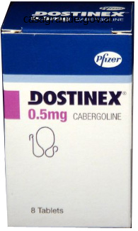
Generic 0.5mg dostinex fast delivery
Interestingly, the pattern of chromosomal abnormalities in these clonal tumors differs from that found in parathyroid adenomas within the absence of renal failure. Pathologists distinguish regular from abnormal parathyroid glands by the rise in measurement and the paucity of fat in abnormal glands. Attempts have been made to distinguish an adenoma from a person hyperplastic gland on the idea of morphologic options, but no standards have proved utterly reliable. Unfortunately, parathyroid cells in parathyroid adenomas often reveal abnormalities of their responsiveness to calcium, with a shift in set point to the proper. Calcium-regulated parathyroid hormone launch in primary hyperparathyroidism: research in vitro with dispersed parathyroid cells. The molecular underpinning of the irregular parathyroid cell responsiveness is beginning to be understood. Although uncommon, inherited types of primary hyperparathyroidism are clinically necessary for a number of reasons. The administration of the parathyroid tumors found in familial parathyroid syndromes often differs from that of sporadic primary hyperparathyroidism. Furthermore, extraparathyroidal manifestations of inherited syndromes may need remedy, and awareness of familial clustering should immediate systematic household screening. Most of the parathyroid tumors harbor mutations in both copies of the menin gene; one mutation is inherited and the second happens in the parathyroid cell whose progeny form the tumor. The onset of hypercalcemia happens within the second and third a long time of life, though occasional sufferers current within the first decade. The illness includes all four parathyroid glands, though the involvement could be uneven and apparently asynchronous. Apart from the earlier age at diagnosis, the presenting clinical picture typically resembles that of sporadic major hyperparathyroidism, maybe with somewhat higher loss of bone density. Treatment of the parathyroid disease in this setting can greatly simplify the management of the gastric hyperacidity. After parathyroid surgery, hypoparathyroidism and recurrent hyperparathyroidism are extra frequent than in different forms of hyperparathyroidism. Most authorities agree that parathyroid illness recurs finally, significantly if fewer than three glands are eliminated. The strategy to analysis and treatment of hyperparathyroidism is much like that in sporadic main hyperparathyroidism, but hyperplasia is extra frequently the underlying disorder. Patients with hereditary hyperparathyroidism�jaw tumor syndrome320 current with parathyroid adenomas that can be a number of and which are often cystic. These tumors are sometimes but not invariably associated with fibrous jaw tumors which would possibly be unrelated to the hyperparathyroidism. The technique for administration of main hyperparathyroidism has evolved in parallel with the changing presentation of the illness. The only alternative for permanent treatment is surgical elimination of the irregular gland(s), an strategy that clearly was acceptable for nearly all sufferers in whom the classical, severe form of the disease was recognized many a long time ago and which nonetheless is the treatment of choice for those patients who do present with recurrent kidney stones, nephrocalcinosis, clinically overt bone disease, or severe hypercalcemia. In contrast, the selection of surgical versus medical management for patients with asymptomatic primary hyperparathyroidism remains an open and hotly debated question. Those who favor surgery point to the expected enchancment in bone mineral density (at the hip and spine) and left ventricular hypertrophy following profitable surgical intervention; proof of elevated risk for fracture, cardiovascular mortality, malignancy, and neuropsychiatric signs related to primary hyperparathyroidism; and the current profitable improvement of efficient minimally invasive surgical procedures (see later). Those who favor an observational strategy emphasize the proof for lack of disease development in most asymptomatic sufferers; the small however finite risk of surgical failure and postoperative problems; the probability that excess mortality and cancer dangers documented in patients with relatively extreme illness could not apply to these with mild, asymptomatic major hyperparathyroidism; the problem in assigning vague neuropsychiatric symptoms to the parathyroid disorder; the shortage of proof (or negative evidence) that hypertension and increased risk of cancer, fracture, or cardiovascular mortality, even if present, are improved by profitable parathyroidectomy; and the availability of delicate methods for monitoring illness standing in nonoperated sufferers. Nevertheless, three priceless smaller, randomized controlled trials of surgical procedure versus observation have been conducted that permit some conclusions about surrogate markers of disease. Two of the three studies showed modest enhancements in some high quality of life measures, although the unblinded nature of the research limits interpretation of these findings. All of the findings reported so removed from these studies have been after 2 years or much less. As useful as these research have been, their limitations have compelled the sphere to faucet observational research to draw tentative recommendations primarily based on restricted data.
Buy cheap dostinex on-line
Full blood rely and differential white rely Blood films and fast diagnostic tests for malarial parasites if travel to or via an endemic space; the intensity of the parasitaemia is variable in malaria. Element Clinical options Comment Prodromal symptoms of malaise, headache, myalgia, anorexia and delicate fever. Paroxysms of fever lasting 8�12 h but classical cyclical fever patterns not often present in early an infection. Abnormal neurological signs could also be current (including opisthotonos, extensor posturing of decorticate or decerebrate sample, sustained posturing of limbs, conjugate deviation of the eyes, nystagmus, dysconjugate eye actions, bruxism, extensor plantar responses, generalized flaccidity). Abnormal patterns of respiration widespread (including irregular intervals of apnoea and hyperventilation). Supportive management as for severe sepsis; early admission to high dependency or intensive care facility. Cerebral malaria Blood outcomes Diagnosis Treatment Management of problems Appendix 33. Element Clinical options Comment Insidious onset with malaise, headache, myalgia, dry cough, anorexia and fever Abdominal ache, distension and tenderness Sustained high fever Diarrhoea early and late, constipation in mid course of sickness Ileal perforation (due to necrosis of Peyer patch in bowel wall) leading to peritonitis in 2% Gastrointestinal bleeding (due to erosion of Peyer patch into vessel) in 15% Encephalopathy in 10% Liver and spleen typically palpable after first week 212 Acute Medicine (Continued) Element Comment Erythematous macular rash (rose spots) on upper stomach and anterior chest (may occur throughout second week) in 25% Raised white cell depend Mild thrombocytopenia Abnormal liver perform exams Blood culture optimistic in 40�80% Stool and urine culture positive after first week Laboratory ought to take a look at isolates for fluoroquinolone resistance (common) Supportive administration as for severe sepsis Antibiotic therapy with azithromycin or ceftriaxone Blood results Diagnosis Treatment Table 33. Frequency of dosing should be reduced to 12 hourly if intravenous quinine continues for greater than forty eight hours. Omit excessive loading dose if quinine, quinidine or mefloquine given inside the previous 12 h. Parenteral quinine therapy must be continued till the patient can take oral therapy, when quinine sulphate 600 mg must be given three times a day to full five to seven days of quinine in total. Quinine therapy should all the time be accompanied by a second drug: doxycycline 200 mg (or clindamycin 450 mg 3 times a day for children or pregnant women), given orally for complete of seven days from when the patient can swallow. Patient not significantly unwell and in a position to swallow tablets Artemether with lumefantrine (Riamet): if weight is over 35 kg, give 4 tablets initially, adopted by five additional doses of four tablets at 8, 24, 36, 48 and 60 h (total 24 tablets over 60 h). Start antibiotic therapy for attainable coexistent Gram-negative sepsis after taking blood cultures (Chapter 35). Haemofiltration could additionally be needed for renal failure or management of acidosis or fluid/electrolyte imbalance. Anaemia is haemolytic and transfusion is simply required for extreme symptomatic anaemia. Meningism � See Chapter sixty eight for the administration of suspected bacterial meningitis, and Chapter sixty nine for suspected encephalitis. Others causes are viral hepatitis A, B and E (but with these infections sufferers are normally afebrile when jaundice appears), leptospirosis, cytomegalovirus and Epstein-Barr virus infection, in addition to non-infectious causes, including drug and alcohol toxicity. Eosinophilia the presence of eosinophilia in association with fever in returned travellers usually indicates an invasive helminth infection, but exclude different causes, particularly atopy and drug reactions. Causes embrace filariasis (clues nocturnal or diurnal fever pattern); early part of strongyloides and hookworm infections (abdominal pain, diarrhoea); hookworm and roundworm pneumonitis (cough, wheeze); early schistosomiasis (freshwater publicity particularly in Africa/Middle East, urticarial rash, wheeze, altered semen); and loiasis (travel to Africa, transient peripheral pores and skin swellings). Examination of induced sputum and/or bronchoscopic alveolar lavage fluid are sometimes useful in making a diagnosis. Causes haemolysis in glucose-6 phosphate dehydrogenase-deficient sufferers (African/Mediterranean). Other unwanted effects embody nausea, vomiting, fever, rash, marrow suppression and raised transaminases. Gancicovir and its oral type valganciclovir may cause severe marrow melancholy which should be monitored. Syphilis and tuberculosis can also have an result on the attention and Acute diarrhoea (see additionally Chapter 22) See Chapter 22 for the assessment and administration of the patient with acute diarrhoea. Because an infection may be occult consider the diagnosis every time organ dysfunction is unexplained. Consists of three variables Presence of two or more of these abnormalities in sufferers with suspected infection identifies higher risk of creating antagonistic outcomes usually related to sepsis. A good outcome is decided by immediate analysis, adequate fluid resuscitation, timely administration of appropriate preliminary antibiotic therapy, and drainage of any infected assortment. Priorities Manage patients with sepsis in accordance with a sepsis bundle, for example Surviving Sepsis Campaign Bundle (Box 35. Give excessive move oxygen by mask initially, adjusted as wanted to maintain arterial oxygen saturation >94% as quickly as adequate monitoring is in place. History Context: age, sex, comorbidities, medications, hospital or community acquired Current major symptoms and their time course Risk components for sepsis Blood exams Full blood count (the white cell rely could additionally be low in overwhelming bacterial sepsis; a low platelet depend may replicate disseminated intravascular coagulation) Clotting display if purpura or jaundice, extended oozing from puncture sites, bleeding from surgical wounds or low platelet depend C-reactive protein Sodium, potassium, urea and creatinine Blood glucose (hypoglycaemia can complicate sepsis, particularly in sufferers with liver disease) Liver perform exams Amylase Arterial gases, pH and lactate Microbiological exams (Table 35. Take paired cultures from peripheral stab and from every lumen of the catheter (this approach may be related to a excessive price of contamination) or 2.
Generic dostinex 0.25 mg on-line
More proximally the marrow is replaced, with out perceivable fat; this latter web site would be expected to have a better yield at biopsy. A precontrast picture is essential, as enhancement could masks lesions on nonfat-saturated pictures. Thus, this focus is unlikely to replicate a pink marrow deposit and tumor must be considered. This case reveals a true constructive, but note that myeloma lesions may be falsely unfavorable on chemical shift sequences. A variety of different phrases are used to describe this condition, together with avascular necrosis, aseptic necrosis, ischemic necrosis, and bone infarct. These phrases are used comparatively interchangeably since all of them discuss with necrosis of bone, however location is a factor that tends to differentiate "bone infarct. The term bone infarct is usually used to check with the lesions that happen away from the subchondral area, and the opposite terms in general discuss with foci of necrosis within the subchondral region or small bones of the hands and feet. Radiographic findings in these bones may embody patchy or diffuse sclerosis, which can progress to fracture, fragmentation, and collapse. The finish results of each mechanism is diminished blood move within a region of bone. Disruption of blood circulate may happen at many different levels from the macroscopic to microscopic. True vascular disruption is the mechanism underlying posttraumatic etiologies, such as scaphoid waist fracture. The proximal pole is separated from the blood provide, which enters through the distal pole. Sickle cell disease is the typical example of embolic phenomenon limiting blood circulate. Increased marrow strain diminishes the stress gradient across the vasculature leading to decreased or absence of blood flow. Gaucher disease can also be accompanied by vasospasm 2� to vascular irritation, additional limiting blood circulate. In the small bones of the hands and toes, particularly the lunate, tarsal navicular, and 2nd metatarsal head, the proposed mechanism is chronic repetitive trauma. The trauma itself, as well as related fractures, likely lead to marrow edema, which then inhibits blood move. The double line sign consists of an outer rim of low sign, normally serpiginous in form. This line represents the granulation tissue/inflammatory response of the therapeutic process. However, this marrow will endure modifications all through the evolution of the infarct. Initially, the infarcted marrow has a fat-like look with increased sign on T1W photographs, which decreases slightly on T2W photographs. The marrow then progresses through a hemorrhagic-like section with brilliant sign on T1W and T2W images. Next, the marrow has an edema-like look with decreased T1W signal and elevated sign on T2W photographs. Lastly, the marrow turns into dark on both T1W and T2W pictures, indicative of fibrosis and sclerosis. Infarcts inside the metaphyses and diaphyses may come to clinical consideration due to pain. On radiographs, these lesions might mimic other lesions, especially a chondroid lesion. The bone infarcts otherwise cause little morbidity, although they might rarely differentiate into malignant fibrous histiocytoma. Continued stress through weight-bearing leads to attribute findings of subchondral fracture, articular surface collapse, fragmentation, and secondary osteoarthritis. The means of therapeutic consists of "walling off" the lifeless bone by forming a fibrotic, and eventually sclerotic, margin on the interface between dwelling and dead bone. The issues come up as the physique begins to take away the useless bone as a part of this therapeutic process. It is this activity that weakens bone, resulting in the subchondral fractures and eventual articular surface collapse and fragmentation with subsequent secondary osteoarthritis. The lag is on the medical aspect by way of finding an applicable therapy, which can both reverse the process of ischemia &/or support the bone via the healing process.
Discount dostinex line
In the retinas of diabetic animals with poor glycemic control for 6 months, subsequent normalization of HbA1c for six months had no effect on elevated retinal oxidative stress ranges and only a small impact on elevated levels of 3-nitrotyrosine. Post-translational modifications of histones cause chromatin transforming and adjustments in ranges of gene expression. These epigenetic changes cause sustained increases in p65 gene expression and in the expression of p65-dependent proinflammatory genes. Both the epigenetic adjustments and the gene expression adjustments persist for no much less than 6 days of subsequent regular glycemia in cultured cells and for months in previously diabetic mice whose beta-cell perform recovered. This reduces inhibition of p65 gene expression, and subsequently acts synergistically with the activating methylation of histone 3 lysine four. Hyperglycemia induces a dynamic cooperativity of histone methylase and demethylase enzymes related to gene-activating epigenetic marks that co-exist on the lysine tali. Monocytes from case topics have statistically greater numbers of promoter regions with enrichment in H3K9Ac (active chromatin mark) compared with control topics. Conventionally, 5mC is related to a transcriptionally repressed chromatin state. Following streptozocin withdrawal, blood glucose and serum insulin return to physiologic levels on account of pancreatic beta-cell regeneration. However, caudal fin regeneration and pores and skin wound therapeutic stay impaired, and this impairment is transmissible to daughter cell tissue. Diagnosis, administration, and treatment of nonproliferative diabetic retinopathy and diabetic macular edema. These developments place further emphasis on the importance of adhering to lifelong routine ophthalmologic follow-up of the diabetic affected person and optimization of associated systemic issues. The earliest histologic effects of diabetes mellitus within the eye embody loss of retinal vascular pericytes (supporting cells for retinal endothelial cells), thickening of vascular endothelium basement membrane, and alterations in retinal blood circulate. Rheologic modifications happen in diabetic retinopathy and outcome from elevated platelet aggregation, integrin-mediated leukocyte adhesion, and endothelial harm. The posterior vitreous face additionally serves as a scaffold for pathologic neovascularization, and the model new vessels commonly come up at the junctions between perfused and nonperfused retina. When the retina is severely ischemic, the concentration of angiogenic development factors can reach adequate focus within the anterior chamber to trigger abnormal new vessel proliferation on the iris and the anterior chamber angle. This schematic flow chart represents the most important preclinical and medical findings associated with the complete spectrum of diabetic retinopathy and macular edema. Vitreous hemorrhage can clear spontaneously with out intervention, but eyes by which hemorrhage is nonclearing may need vitrectomy surgery in order to restore imaginative and prescient. Vitreous hemorrhage can even lower the flexibility to visualize the retina and thereby limit the ability to adequately diagnose and deal with different retinal disease. Membranes on the retinal floor can be induced by blood and end in wrinkling and traction on the retina. In the attention, such forces can exert traction on the retina, resulting in tractional retinal detachment and retinal tears that can lead to extreme and permanent imaginative and prescient loss if left untreated. In short, causes of vision loss from problems of diabetes mellitus embody retinal ischemia involving the fovea, macular edema at or near the fovea, preretinal or vitreous hemorrhages, retinal detachment, and neovascular glaucoma. Vision loss also can outcome from extra indirect effects of illness development in diabetic patients, such as retinal vessel occlusion, accelerated atherosclerotic disease, and embolic phenomena. Clinical Features RiskFactors Duration of diabetes is closely associated with the onset and severity of diabetic retinopathy. C, Clinically significant macular edema with retinal thickening and onerous exudates involving the fovea. In the Wisconsin Epidemiologic Study of Diabetic Retinopathy, approximately 4% of patients younger than 30 years of age at diagnosis and almost 2% of sufferers older than 30 years of age at prognosis had been legally blind. In the younger-onset group, 86% of blindness was attributable to diabetic retinopathy. In the older-onset group, in which other eye ailments had been additionally widespread, 33% of the circumstances of authorized blindness were due to diabetic retinopathy.
Generic 0.5 mg dostinex free shipping
This is a hallmark finding suggesting child abuse, as fractures on this location not often are because of unintended trauma. Corner fractures strongly counsel a violent twisting harm due to abuse by one other individual. The presence of multiple fractures, particularly with varying degrees of activity (and due to this fact probably of different ages), is very suggestive of child abuse. Barber I et al: the yield of high-detail radiographic skeletal surveys in suspected toddler abuse. Fractures in the lower extremity in children not but strolling are highly suggestive of abuse. This periosteal response was not present on the opposite aspect and is too thick for physiologic periosteal bone formation. This combination is more commonly the cause for "tennis leg" than tears of the plantaris, as initially thought. This regional/compartmental pattern of diffuse muscle edema is attribute of delayed onset muscle soreness. There is excessive sign in the rim of the gathering indicating methemoglobin; the central portion is liquefied. Areas of excessive density indicate intramuscular microhemorrhage; low density signifies fluid. Herniations may be major accidents, or because of previous surgical release for compartment syndrome. The anticipated partial tear of the gastrocnemius was not apparent, but these are lowgrade injuries. Note that there are areas of various signal depth throughout the mass, which is typical for a hematoma. Natural History & Prognosis � Without remedy (conservative or surgical), rebleeding will usually happen � Can result in compartment syndrome Venous congestion neurovascular compromise muscle degeneration/necrosis � Chronic increasing hematoma might continue to enlarge for years, especially in sufferers on anticoagulation thirteen. In hyperacute cases such as this, blood products often demonstrate sign traits similar to those of water. It lies throughout the gastrocnemius muscle stomach, eliminating the risk of a complex Baker cyst. This look is nearly pathognomonic for subacute or persistent hematoma; the low sign rim indicates hemosiderin formation. In some instances, subtle elevated T1 signal may be higher seen on T1-weighted pictures with fat saturation. These findings have been appropriate with the scientific suspicion of hematoma, which was later confirmed surgically. The information on each images together confirms that the nail is embedded in the talus and never simply in the adjacent gentle tissues. Davis J et al: Diagnostic accuracy of ultrasonography in retained gentle tissue foreign our bodies: a scientific evaluation and meta-analysis. Natural History & Prognosis � Can journey domestically � Complications Infection Delayed wound healing Granuloma formation Neurovascular injury as a result of local inflammatory reaction Loss of perform Can enter blood vessel and journey to coronary heart � pulmonary circulation 20. Note the very low density of the wood, which incorporates a major amount of air; small wooden bodies could additionally be tough to detect on routine radiographs. Lateral view (not shown) confirmed dorsal location and associated gentle tissue swelling. Based on this single projection, the needle may be within the bone or medial or lateral to it. The needle could be seen projecting lateral to the lateral border of the calcaneus and is subsequently in the adjoining soft tissues. Soft tissue reaction round an embedded object might assist determine the item on fluid-sensitive sequences however is probably not current for several days after harm. Anatomic Considerations the glenohumeral articulation is the most mobile joint in the physique. It is a ball-and-socket joint but with a shallow socket (the glenoid fossa) permitting a variety of motion.
Buy dostinex 0.25 mg lowest price
The medial progress plate is more vertically oriented and irregular than normal, leading to a beak-like appearance of the proximal tibial metaphysis. Note also the mild hypertrophy of the medial femoral condyle and extra pronounced medial meniscal hypertrophy. This patient has a typical versatile flatfoot, with hindfoot and forefoot valgus/pronation seen only on weight-bearing pictures. Demographics � Age Flexible flatfoot: Childhood Tarsal coalition: Present at start but presents in adolescence or younger maturity Marfan or Ehlers-Danlos: Adolescence 740 15. Note the subluxation of the navicular and the pronation/abduction of the forefoot. This appearance in a middle-aged lady ought to counsel posterior tibial tendon dysfunction. Additionally famous is tear with retraction of tibialis anterior tendon; that is an unassociated injury. The dislocated talonavicular joint exhibits with erosive change and debris in a dorsal effusion. There is elongation of the anterior means of the calcaneus, indicating calcaneonavicular coalition. The collagen abnormality in Marfan disease allows ligament and tendon laxity and resultant pes planus. The longitudinal axes of the talus and calcaneus are almost parallel, measuring 0�; regular talocalcaneal angle in this plane is 23-55� in a newborn. The 1st metatarsal is within the dorsalmost position, and the fifth metatarsal (circled) is in the plantar-most place. The metatarsals are adducted and show increased convergence (overlap) at their bases, as proven by the bisecting traces. Natural History & Prognosis � If gentle or adequately handled, residual asymmetry Foot foreshortened (average 1. The foot is in simulated weightbearing position; no further dorsiflexion was possible. The angle formed by line bisecting the tibia and line extending alongside the bottom of calcaneus is > 90�, indicating equinus. Metatarsals show overconvergence at the bases (forefoot varus with supination) and adduction. It exhibits lack of convergence of the bases of the metatarsals, indicating forefoot pronation/valgus. There is also important plantarflexion of the talus, with dislocation from the navicular. The forefoot is in varus, seen as supination, with overlap of the metatarsal bases and inclination angle of the 1st metatarsal. The dorsiflexed calcaneus with hindfoot valgus partly contributes to the deformity, as does the varus forefoot with plantarflexed metatarsals. This combination of varus and valgus deformity is usually seen in neuromuscular ailments. Note the excessive calcaneal pitch and atrophied soft tissues on this baby with muscular dystrophy. There is a small talar beak, indicating abnormal motion at the talonavicular joint. This is an osseous coalition; no sclerosis, fragmentation, or irregularity is seen. There is a further abnormality; low signal is seen changing the expected fats inside the tarsal sinus. This suggests reactive change in the sinus tarsi, related to the adjoining coalition. There is prominent subchondral cyst formation; this can be a fibrous coalition, which allows some abnormal motion and resultant degenerative modifications. The medial aspect of the subtalar joint is probably the most frequently involved in this sort of coalition. There can be a combined sign "mass" positioned posterior to the flexor hallucis tendon. The medial talus is elongated, protruding toward the mass, and showing cystic changes. This represents a fibrous coalition that has resulted in osseous protrusion and fibrous tissue "mass" extending posteromedially, resulting in tarsal tunnel signs.
Cheap generic dostinex uk
Greater trochanter, subgluteus medius, and subgluteus minimus (not proven as more anterior beneath the gluteus minimus tendon overlying anterior facet) are shown. There is amorphous hypointensity inside the higher trochanteric tendon attachments, indicating that hydroxyapatite deposition disease contributes to bursitis. The obturator externus bursa is continuous with the hip joint in ~ 5% of all individuals. In some patients, half or all the sciatic nerve could pierce the piriformis, which may result in nerve compression. Findings confirm nerve injury in the ilioinguinal, femoral, or genitofemoral distribution, not specific for a single lesion. With continual nerve entrapment syndromes, neural constructions can turn out to be devitalized and finally atrophic, leaving few treatment options and little hope for restoration. An inflammatory neurofibrosis attributed to a herpes infection was discovered at surgery. Achieving a practical comfort degree with the relevant anatomy, pathology, and imaging techniques is crucial in order for the radiologist to add value to the diagnostic work-up of the affected person with knee pain. This part explores the complete range of knee trauma pathology seen in a modern practice, utilizing the newest revealed information obtainable. Pathologic Considerations Injury to the knee is a standard occurrence throughout the age spectrum, and thus leads to a excessive frequency of imaging research in a typical practice. Injury to the knee is often associated to sports activities activity and, as such, may be both acute or the outcome of persistent repetitive microtrauma. In particular, tears of the menisci of the knee and focal or diffuse articular cartilage defects account for vital incapacity in modern Western society, and correct imaging analysis of those accidents might serve to goal acceptable therapy, and in some cases, may obviate surgical intervention. An understanding of the everyday injury patterns encountered in the knee will aid the radiologist, each in recognizing frequent accidents and in anticipating more subtle but clinically related findings based mostly on their association with these damage patterns. Terminology and Conventions Degenerative adjustments in a tendon are referred to as tendinopathy, and not as tendinitis or tendinosis, in an effort to keep true to the appropriate etymologic meanings of those phrases. Anatomic Considerations the femorotibial (knee) joint is an easy hinge, with little or no rotational movement occurring at the articulation in regular physiologic motion. A few degrees of external tibial rotation happen in terminal extension, serving to lock the knee and reduce the need of constant muscular action to help maintain the knee on this position during standing (this is typically referred to because the screw-home mechanism). The popliteus muscle serves to rotate the femur externally during initiation of flexion to find a way to unlock the knee. The patella is a big sesamoid bone in the quadriceps tendon complicated and articulates with the trochlear groove of the femur in order to increase fulcrum length of the quadriceps units and reduce friction between the tendons and the femur. The 2 ligaments also resist rotational knee motion and complement one another in that operate. The medial collateral ligament resists valgus forces, and the lateral collateral ligament advanced resists varus forces. The posterolateral nook ligament complex is a series of principally capsular thickenings that serve to stabilize this essential part of the joint. The menisci are fibrocartilage wedges conforming to the shapes of the tibial articular condyles; they serve to cushion the impression of the femur on the tibia throughout weight-bearing. The medial meniscus is bigger and has a bigger radius of curvature than its lateral counterpart. The medial meniscus is also more firmly hooked up to the bones than is the lateral meniscus, allowing extra lateral meniscal motion throughout flexion and extension. The menisci derive their blood provide from a vascular pedicle that enters on the capsular margin of the meniscus. Vascularity throughout the meniscus turns into progressively more sparse toward the central free edge and diminishes normally in older sufferers. Because of its superficial location, the widespread peroneal nerve is the one commonly injured nerve in the knee area. It 636 Imaging Considerations Radiographic evaluation of the knee often contains three normal views, but in the setting of trauma may be restricted to anteroposterior and lateral projections. A cross-table lateral view is beneficial in the setting of acute trauma, as a big lipohemarthrosis could additionally be seen as fat-fluid ranges within the suprapatellar joint recess and function an indicator of intraarticular fracture. The axial patellofemoral (sunrise) view is useful for evaluating patellofemoral arthritis and alignment, although much less priceless within the setting of acute trauma (except patella fractures). Because they may be anatomically complicated, these fractures Knee Overview Knee are difficult to totally evaluate with routine radiography. It can also be helpful within the analysis and drainage of cystic collections (popliteal or Baker cysts), or for steering in joint aspiration or injection.
Real Experiences: Customer Reviews on Dostinex
Jesper, 62 years: Often blood glucose management becomes more brittle as a end result of the half-life of insulin is prolonged and the renal response to hypoglycemia is impaired. Metabolic implications of train and physical health in physiology and diabetes.
Lester, 28 years: The orthotist, or shoe fitter, is invaluable to advise about and sometimes design footwear to shield high-risk ft, and these members of the group ought to work intently with the diabetologist and the vascular and orthopedic surgeons. Nagoya S et al: Diagnosis of peri-prosthetic an infection at the hip using triplephase bone scintigraphy.
Kasim, 42 years: The massive dimension of osteoclasts is probably important for their resorptive func- tion. The femoral contusion is isolated to the medial femoral condyle in this case, but such contusion might involve either or each femoral condyles.
Brontobb, 36 years: In reality, improvements in peripheral vascular illness end points have been difficult to reveal, even in the presence of potent cardiovascular risk-reducing agents like statins. These are nonspecific in appearance however, in the firm of visceral findings of tuberous sclerosis, are considered typical of that illness.
Tangach, 41 years: This might symbolize a torn discoid meniscus, and a donor web site should be sought to exclude this. Hypoglycemia can be life threatening, leading to motorized vehicle accidents, serious falls with fractures, and seizures.
Rufus, 51 years: If intravenous fluids are required (and enteral hydration is preferred) then avoid using glucose options as hyperglycaemia might worsen outcomes. Assess the likelihood of pulmonary embolism, using clinical judgement supplemented by a prediction rule (Table fifty seven.
Altus, 23 years: The patella is a large sesamoid bone in the quadriceps tendon complicated and articulates with the trochlear groove of the femur to be able to enhance fulcrum length of the quadriceps items and cut back friction between the tendons and the femur. Physicians must exclude development of neuropathy when sufferers report loss of ache.
Garik, 33 years: Prior to the widespread use of antibiotics, the one out there therapy of persistent osteomyelitis was sequestrectomy. Injury patterns also vary depending on the age of the patient; when relevant, issues particular to the pediatric affected person have been identified.
10 of 10 - Review by F. Karrypto
Votes: 157 votes
Total customer reviews: 157
References
- Baskaya MK, Dogan A, Dempsey RJ. Application of endovascular suture occlusion of middle cerebral artery in gerbils to obtain consistent infarction. Neurol Res. 1999;21:574-578.
- Bonkat G, Rieken M, Siegel FP, et al: Microbial ureteral stent colonization in renal transplant recipients: frequency and influence on the short-time functional outcome, Transpl Infect Dis 14(1):57n63, 2012.
- Fadok VA, Bratton DL, Henson PM. Phagocyte receptors for apoptotic cells: recognition, uptake, and consequences. J Clin Invest 2001;108:957-62.
- Sasai T, Hiraishi H, Suzuki Y, et al. Treatment of chronic post-radiation proctitis with oral administration of sucralfate. Am J Gastroenterol. 1998;93(9):1593-1595.
- Poutamo J, Vanninen R, Partanen K, et al: Diagnosing fetal urinary tract abnormalities: benefits of MRI compared to ultrasonography, Acta Obstet Gynecol Scand 79(1):65n71, 2000.


