B Douglas Smith, M.D.
- Co-director, Clinical Research Operations for the Division of Hematologic Malignancies
- Professor of Oncology

https://www.hopkinsmedicine.org/profiles/results/directory/profile/0008136/b-smith
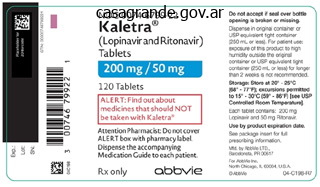
Kaletra dosages: 250 mg
Kaletra packs: 60 pills, 120 pills, 180 pills, 240 pills, 300 pills, 360 pills
Buy kaletra in united states online
Describe the radiographic options of an inflammatory arthritis (synovial-based diseases). The irritation also leads to hyperemia, which, coupled with the inflammatory mediators released (such as prostaglandin E2), causes periarticular (juxtaarticular) osteopenia. With chronicity, inflammatory arthritis may result in more diffuse osteoporosis (due to disuse and other factors) of the joints due to ache. As the inflammation leads to synovial hypertrophy and pannus formation, the pannus erodes into the bone. Cartilage destruction results either by enzymatic action of the inflamed synovium and/or by interference with normal cartilage vitamin. Owing to its generalized nature, this cartilage destruction is radiographically seen as uniform or symmetric, diffuse joint-space narrowing observed best in weight-bearing joints. It is essential to remember that some findings of degenerative arthritis may be superimposed on those of an inflammatory nature, significantly in long-standing cases. In synovial articulations, hyaline articular cartilage covers the ends of each bones. The articular capsule envelopes the joint cavity and is composed of an outer fibrous capsule and a thin inside synovial membrane. Radiograph of a hand displaying periarticular osteopenia and bony erosions (arrows) suitable with inflammatory arthritis in this patient with rheumatoid arthritis. Small black arrows level to "bare areas," the place the bone is exposed to synovium with out protecting cartilage covering. These "naked areas" are where you need to search for the earliest evidence of erosions. With development of the illness, the pannus proliferates to cowl the cartilage surfaces, resulting in cartilage destruction (joint-space narrowing) and more diffuse bony erosions. List the rheumatic illness categories that typically trigger radiographic features of inflammatory arthritis. Describe the radiographic features of noninflammatory, degenerative arthritis (cartilage-based diseases). Nonuniform lack of cartilage (focal joint-space narrowing in area of maximal stress in weight-bearing joints). However, in contrast to uniform, diffuse narrowing seen with inflammatory arthritides, the noninflammatory, degenerative arthritides tend to have nonuniform, focal joint-space narrowing, being most pronounced in the area of the joint the place stresses are extra concentrated. Following cartilage loss, subchondral bone turns into sclerotic or eburnated, owing to trabecular compression and reactive bone deposition. With denudation of the cartilage, synovial fluid can be forced into the underlying bone, forming subchondral cysts or geodes with sclerotic margins. As an attempted reparative process, the remaining cartilage undergoes endochondral ossification to develop osteophytes. Such osteophytes generally happen first at margins or nonstressed features of the joint. List the rheumatic illness classes that sometimes cause radiographic options of noninflammatory arthritis. Knee radiograph demonstrating osteophytes (arrows) and medial joint-space narrowing according to degenerative arthritis. Hand radiograph with degenerative options including joint-space narrowing and osteophyte formation. What are the typical sites of joint involvement in primary (idiopathic) osteoarthritis compared with secondary causes of noninflammatory, degenerative arthritis? Primary (idiopathic) osteoarthritis can cause noninflammatory, degenerative arthritic modifications within the following joints: · Hands. Secondary causes of degenerative arthritis may find yourself in noninflammatory, degenerative modifications in any joint (not just those for primary disease). Consequently, if a affected person has degenerative adjustments in any of the next joints, you should consider secondary causes of osteoarthritis: · Hands.

Purchase kaletra 250mg fast delivery
Palpation for synovitis is finest accomplished over the anterior (not lateral) side of the joint. When the ankle is at the normal place of relaxation (right angle between foot and leg), the ankle usually has 20 levels of dorsiflexion and forty five levels of plantar flexion. Pain worse on first getting away from bed in morning with weight stretching plantar fascia. The foot is often uncared for in the bodily examination but can be a supply of decrease extremity pain. This is called a "gait of ache" and often signifies ache in knee, ankle, or foot. This "gait of weak point" is just like the coxalgic gait in appearance however is because of weakness and not pain. BiBliography American College of Rheumatology Ad Hoc Committee on Clinical Guidelines: Guidelines for the preliminary evaluation of the grownup affected person with acute musculoskeletal signs, Arthritis Rheum 39:1, 1996. Acute-phase reactants are a heterogeneous group of proteins (fibrinogen, haptoglobin, C-reactive protein, alpha-1-antitrypsin, and others) which are synthesized within the liver in response to irritation. Aging, female sex, obesity, being pregnant, and presumably race are noninflammatory conditions that may elevate the sedimentation price. Complete history and physical examination and routine screening laboratories (complete blood rely, chemistries, liver enzymes, urinalysis). It is present in trace concentrations within the plasma of all humans, and it has been extremely conserved over hundreds of hundreds of thousands of years of evolution. Although its exact perform is unknown, it reveals important recognition and activation properties. In the absence of inflammatory stimuli, it falls rapidly, with a half-life of about 18 hours. Both checks measure elements of the acute-phase response and are helpful in measuring generalized irritation. Although serum protein electrophoresis is the most expensive test (Medicare cost=$31. Inflammation is adopted by characteristic protein alterations which are reflected on highresolution electrophoresis. The typical sample consists of increases in immunoglobulins in addition to increases in the -1 zone. Decreases (negative acute section reactants) are seen in prealbumin, albumin, and the zone (transferrin). Cells are visualized via a fluorescence microscope to detect nuclear fluorescence. Each laboratory should decide the extent that it considers positive, and this stage could range considerably amongst labs. Certain patterns of fluorescence are related to sure nuclear antigens are related to particular illnesses (Box 6-1). Patterns of staining provide a clue to the class of nuclear antigens involved and are dependent upon the kind of substrate used, and to a sure extent, the experience of the technician. In some diseases, antibodies towards cytoplasmic antigens may be extra helpful diagnostically than antibodies towards nuclear antigens. Consequently, the particular anticytoplasmic antibody should be ordered when these illnesses are suspected. It capabilities as an E3 ubiquitin ligase that adds ubiquitin to a number of proteins involved in the inflammatory and immune response resulting in their accelerated degradation. Other antigen targets embody elastase, cathepsin G, lactoferrin, lysozyme, and azurocidin. Usually seen in patients with inflammatory bowel illness, connective tissue ailments, or autoimmune hepatitis. Decreased production, owing to either a hereditary deficiency or liver disease (complement components are synthesized in the liver).
Order 250 mg kaletra otc
Plasma exchange is often utilized in mixture with corticosteroids and/or cytotoxic therapy to lower the chance of a rebound flare of the underlying immunologic illness as quickly as the pheresis is stopped. Most plasma change protocols take away 2 to 4 L (40 mL/kg = 1 plasma volume) of plasma over a 2-hour period daily. Replacement fluid is mostly albuminsaline or another protein-containing solution. Discuss the usage of high-dose immunoablative remedy with autologous hematopoietic stem cell transplantation for the treatment of severe autoimmune illness. Some sufferers can also obtain lymphoablative antibodies or total body irradiation to eradicate residual autoreactive cells. This stem cell transplantation strategy allows the patient to reconstitute their immune system without redeveloping their autoimmune disease or developing graft versus host disease. This process is most often used for treatment-resistant systemic lupus erythematosus, systemic sclerosis, and a quantity of sclerosis with varying success charges and a mortality rate as excessive as 8% at a value of up to $100,000. Other immunoablative and/or transplantation methods are also being investigated. BiBliography Ballow M: the IgG molecule as a organic immune response modifier: mechanisms of action of intravenous immune serum globulin in autoimmune and inflammatory problems, J Allergy Clin Immunol 127:315323, 2011. Blumenfeld Z, Shapiro D, Shteinberg M, et al: Preservation of fertility and ovarian function and minimizing gonadotoxicity in young ladies with systemic lupus erythematosus handled by chemotherapy, Lupus 9:401405, 2000. Haubitz M, Bohnenstengel F, Brunkhorst R, et al: Cyclophosphamide pharmakokinetics and dose changes in sufferers with renal insufficiency, Kidney Int sixty one:14951501, 2002. Takada K, Arefayene M, Desta Z, et al: Cytochrome P450 pharmacogenetics as a predictor of toxicity and scientific response to pulse cyclophosphamide in lupus nephritis, Arthritis Rheum 50:22022210, 2004. Tumor necrosis issue inhibitors are more practical when combined with methotrexate. Risk of hepatitis B reactivation and mycobacterial infections are elevated in sufferers on biologics. What biologic agents are presently obtainable to be used in the treatment of inflammatory rheumatic diseases? Cannot bind to Fc receptors, fix complement, or cross placenta as a outcome of not having a practical Fc fragment. The pegylation delays clearance and will help localize the molecule to acidic, inflammatory sites. Rarely enhance dose higher than 5 mg/kg each four weeks due to an infection and malignancy considerations. Most patients will obtain their maximum enchancment inside three months, though some proceed to enhance with continued use. If issues persist, lyophilized etanercept or certilizumab pegol can be used which appear to have fewer injection site reactions. Infections are inclined to be pneumonias which happen more commonly throughout the first 6 months of use. The threat of an infection is lowered if surgical procedure is performed after waiting a minimum of three half-lives from the last dose (9 days after etanercept; four to 5 weeks after infliximab and adalimumab; 6 weeks after golimumab or certilizumab). Owing to lengthy half-life and blood levels, infliximab could trigger more of these infections than subcutaneous formulations. In over 50% of instances the reactivation is at a site other than the lung (lymph nodes commonly). Patients with earlier or current exposure to endemic fungi (Histoplasmosis, Coccidioidomycosis, others) have to be evaluated for these infections if they develop a febrile sickness. Studies range and state that the relative threat may (<5Ч relative risk) or may not be elevated for lymphoma. Many consultants suggest not beginning these agents till a patient is most cancers free for five years. Demyelinating syndromes Brain demyelination (multiple sclerosis-like), optic neuritis, GuillainBarrй syndrome, polyradiculopathy, and peripheral demyelinating neuropathy have been reported rarely. Some consultants advocate brain magnetic resonance imaging in sufferers with a robust family historical past of demyelinating illness to look for occult lesions. Hematologic Neutropenia, thrombocytopenia, and pancytopenia have hardly ever been reported.
Order genuine kaletra online
As much as 35% primipara women have proven to have sustained occult sphincter harm as seen on anoendosonogram. Second-degree lacerations should always be sutured fastidiously instantly after supply. The pelvic floor is weakened unless the injury to the muscular tissues of the perineal physique is efficiently repaired. If the decussating fibres of the levator ani muscular tissues are torn by way of, the hiatus urogenitalis becomes patulous and prolapse of the vagina and the uterus is likely to develop, until these lacerations are sutured immediately after supply. With intensive second-degree tears, the patient should be given an area, regional pudendal block or common anaesthetic, placed in the lithotomy place, and the torn muscles of the perineum recognized and sutured together with catgut. The torn edges of the vagina and the skin of the perineum should then be sutured together with catgut. The important a part of the after therapy of perineal lacerations consists in keeping the perineum clean. The wound ought to be cleaned with an antiseptic resolution similar to Betadine after micturition and defaecation. Amongst the predisposing causes of complete tear of the perineum are forceps delivery in the persistent occipitoposterior positions, and extraction of the after coming head in breech presentation. A properly carried out episiotomy will very largely get rid of the risk of a third- and fourth-degree tear. This kind of tear is extra common with median episiotomy than mediolateral episiotomy. Complete tear of the perineum should be repaired as quickly as attainable after the delivery. The operation should be undertaken beneath anaesthesia with the affected person mendacity in the lithotomy position in good gentle and with good help. The quick restore of a whole tear of the perineum is a comparatively easy procedure, for the reason that muscle tissue of the perineal physique, although torn, may be distinguished without much problem. The surrounding skin is first cleaned and the operation area isolated with sterile towels. A sterile pack is placed in the vagina and the bounds of the laceration defined with tissue forceps. A few Lembert sutures are then introduced to invaginate the torn edges of the bowel wall. The muscles of the perineal body are now sutured together, and each effort must be made to get hold of exact anatomical reposition. Particular consideration must be paid to the sphincter ani muscle, and at least two Vicryl sutures must be used to draw the minimize edges collectively. The tears within the vaginal wall and within the skin of the perineum at the moment are repaired with interrupted catgut sutures. Care must be taken to avoid tying the sutures too tightly; otherwise, oedema of the perineum will lead to extreme ache and cause the stitches to cut through. The end outcomes are sometimes functionally good despite the preliminary breakdown of the suture line. The bowels should be confined for no much less than 5 days, solid meals withheld and intestinal antiseptics given, together with stool softeners. Lately, instead of end-to-end suturing of the torn sphincter muscular tissues, overlap method is really helpful to yield a stronger sphincteric control. The purple glistening mucous membrane of the anal canal and rectum protrudes and fuse instantly with the vaginal wall without any of the perineal tissues intervening. Behind the anus are the radial folds in the skin which are corrugated by the underlying contracted subcutaneous sphincter. The external sphincter is just current posteriorly and the absence of the sphincteric grip is appreciated by inserting a finger into the anus. One of probably the most interesting options of the complete tear of the perineum is that it is extremely rarely if ever related to prolapse, though the decussating fibres of the levator ani muscular tissues have been torn through. The cause is that the patient repeatedly attracts together the 2 levator ani muscles in an effort to close the bowel in order that by fixed use the tone of the muscle tissue turns into exceptionally good. This firmness and good development of the levator muscular tissues is found on scientific examination when the levator muscular tissues are palpated. The technical difficulties are much higher in old instances than in these operated upon instantly after supply. The optimum time for operation within the case of old tears is 36 months after delivery.
Cheap kaletra 250mg without a prescription
Imiquimod cream applied 3 times every week for 4 months cures 75% instances, however recurrence occurs in 15% instances. Complications Known issues include encephalitis, urinary tract involvement inflicting retention of urine, extreme pain or both. The prevalence of the illness has reached epidemic proportions in the developed international locations of the world. The affected person typically complains of constitutional signs such as malaise, fever and vulval paraesthesia followed by appearance of vesicles on the vulva leading to ulcers, that are shallow and painful. Viral shedding, nevertheless, tends to proceed for weeks after the appearance of lesions. Prodromal signs of burning or itching within the affected space typically precede the attacks. About 50% of the affected women experience their first recurrence inside 6 months and have on a mean about four recurrences throughout the first 12 months; thereafter, the episodes of recurrences are inclined to happen at variable intervals. Latent herpes virus residing within the dorsal root ganglia of S2, S3 and S4 could get reactivated every time the immune system will get compromised as seen during being pregnant or any other immunocompromised state. In severe cases, administer acyclovir 5 mg/kg body weight intravenously every eight h for five days. Treat major outbreaks: Prescribe oral 200 mg acyclovir five occasions day by day for 5 days. Local utility of acyclovir cream supplies aid and accelerates therapeutic of local lesions. Valacyclovir 500 mg bd or famciclovir 125 250 mg bd can be efficient, given for 7 days. Counselling: the couple is advised to abstain from intercourse from the time of experiencing prodromal symptoms until whole re-epithelialization of the lesions takes place. Chapter eleven · Sexually Transmitted Diseases n 159 Caesarean section is beneficial within the presence of active an infection, to avoid neonatal an infection. Clinical Features It begins as a painless nodule which later ulcerates to form a number of beefy purple painless ulcers that tend to coalesce, the vulva is progressively destroyed and minimal adenopathy could occur. Sexual transmission: In girls, the organism is carried by lymphatic drainage from the genital lesion to the perirectal, both inguinal and pelvic lymph nodes. Rectal involvement is common in females and happens by contiguous unfold from the perirectal nodes leading to proctocolitis and rectal strictures formation. The drainage is primarily to the inguinal nodes leading to bubo formation; this will likely burst, ulcerate or cause sinus. Diagnosis Microscopic examination of smears from the lesion/biopsy specimens reveals pathognomonic intracytoplasmic Donovan bodies and clusters of micro organism with a bipolar (safety-pin) look (Gram negative). Treatment n Clinical Features the lesion begins as painless vesicopustular eruption that heals spontaneously. After some weeks, the sequelae of lymphatic spread begin with hardly any medical manifestations. Investigations the Frei check based mostly on delayed pores and skin hypersensitivity to the antigen turns into optimistic 28 weeks after major an infection. It contains the following: (a) proctitis, (b) severe stricture formation leading to intestinal obstruction, (c) rectovaginal fistulae following stricture formation and (d) vulvar most cancers. Treatment Treatment with tetracyclines 500 mg 6 h, doxycycline a hundred mg bid orally for 3 weeks or sulphonamides or erythromycin 500 mg orally each 6 h every day for 36 weeks are equally efficient in eradicating the disease. Other antibiotics are amoxycillin 500 mg tid for 7 days and azithromycin 1 g single dose. Mycoplasma Genitalium Mycoplasma genitalium, first discovered in 1983, is an intracellular organism lacking cell wall, not stained by Gram stain. The traditional lesion designated as the chancre appears within 990 days from the first publicity. Onset of systemic manifestations includes symptoms such as malaise, headache, lack of appetite, sore throat and the looks of a generalized symmetric, asymptomatic maculopapular rash on the palms and soles of the ft.
Syndromes
- Teach children the proper names of body parts.
- Rash
- Tumors
- Seizure
- D2 (ergocalciferol)
- Unusual bruising and scarring patterns can also be caused by folk medicine or Oriental medicine practices such as coin rubbing, cupping, and burning herbs on the skin over acupuncture points (called moxibustion). The doctor should always ask about alternative healing practices.
- Contact your doctor if you have a cold, flu, fever, herpes breakout, or any other illness.
- Headache
- Joint pain
- Foods: Foods passed through breast milk may affect your child. If you are breastfeeding, avoid stimulants such as caffeine and chocolate. Try to avoid dairy products and nuts for a few weeks, as these may be causing allergic reactions in the baby. People often hear that breastfeeding moms should avoid broccoli, cabbage, beans, and other gas-producing foods. However, there is not much evidence that these foods are a factor.
Generic kaletra 250 mg mastercard
Patients with absent pus at sites of an infection and poor wound therapeutic have defects in neutrophil numbers, adhesion, or chemotaxis. Patients with neutrophil defects of microbial killing current with lymphadenitis and visceral or perirectal abscesses with granuloma formation brought on by low-virulence gram-negative organisms such as Escherichia coli, Serratia, and Klebsiella. Other sufferers will have gingivitis and pores and skin infections or furunculosis with Staphylococcus or Pseudomonas species. Standard neutrophil tests include evaluation of cell number, examination for big granules on a peripheral smear (ChйdiakHigashi syndrome), and measurement of oxidative burst (chronic granulomatous disease). The flow cytometry dihydrorhodamine 123 assay is best than the nitroblue tetrazolium test for assessing neutrophil oxidative burst. Deficiencies of elements of the membrane assault advanced (C5 to C9) or different complement pathway elements are related to recurrent Neisseria infections (both N. Patients with recurrent bouts of neisserial infections, significantly when systemic, should be evaluated for the presence of a complement deficiency. Use of prophylactic antibiotics is controversial due to the risk of causing antibiotic-resistant strains of Neisseria. Complement element deficiencies of the classical pathway have an autosomal recessive pattern of inheritance. What organisms are answerable for septic arthritis in patients with hypogammaglobulinemia as a outcome of primary B-cell immunodeficiency? These patients are susceptible to septic arthritis attributable to the usual organisms encountered in B-cell immunodeficiency states: S. In addition to these typical infectious brokers, sufferers are additionally prone to joint infections with Ureaplasma urealyticum and other Mycoplasma organisms. This ends in characteristic mobile abnormalities: maturation failure for the B cell line and the absence of B cells. Arthritis happens in approximately 20% of sufferers, with half of these circumstances attributable to an infection with the typical pyogenic micro organism. In addition, sufferers appear weak to infections with enterovirus and Mycoplasma Table 58-3). Rheumatologic Manifestations of X-Linked Agammaglobulinemia Septic arthritis Extracellular, encapsulated bacteria (S. The deficiency is characterized by very low ranges (<5 mg/dL) of serum and secretory IgA, accompanied by normal serum ranges of IgG and IgM. Some cases could additionally be acquired later in life, typically associated with drug therapy or viral infection, and are sometimes transient. List the autoantibodies seen in patients with selective IgA deficiency without clinically expressed autoimmune illness. The presence of autoantibodies in the absence of clinically expressed autoimmune disease generally occurs in IgA deficiency. What systemic and organ-specific autoimmune ailments are related to selective IgA deficiency? Table 58-4 lists the autoimmune illnesses related to selective IgA deficiency. Other conditions have been famous in case stories, however their true affiliation with IgA deficiency stays to be proved. Rheumatologic Manifestations of Common Variable Immunodeficiency Septic arthritis Extracellular, and encapsulated micro organism (S. Autoantibody presence in the absence of clinically expressed autoimmune disease occurs less often than in selective IgA deficiency. Characteristics embrace involvement of the big and medium joints, with sparing of the small joints of the arms and feet. This type of arthropathy usually responds to therapy with intravenous gammaglobulin. The hyper-IgM immunodeficiency syndrome (type 1) is characterized by extremely low levels of IgG, IgA, and IgE, and either a normal or markedly elevated concentration of polyclonal IgM. Patients develop each recurrent pyogenic infections and opportunistic infections corresponding to P. This lack of B-cell signaling by T cells leads to a failure of B cells to bear isotype switching, so that they produce solely IgM. Two other types of this syndrome (types 2 and 3) have totally different genetic defects and inheritance patterns.
Purchase kaletra 250mg with amex
Typically, morning stiffness is absent or of brief duration (<1 hour), allowing differentiation from inflammatory monoarticular arthritides. Direct results embody elevated apoptosis of osteoblasts, osteocytes, and endothelial cells. Indirect results include elevated hypercoagulability, decreased angiogenesis, and modulated native vasoactive amine manufacturing, which may contribute to the ischemia. Finally, elevated intraosseous pressure as a result of adipogenesis and fat hypertrophy in the bone marrow can decrease blood circulate to the area. It is estimated that a total of one hundred fifty L of 100% alcohol at a fee of four hundred mL/week. Extensive evaluation helps that many of these sufferers may have a hypercoagulable state as evidenced by elevated lipoprotein(a), low tissue plasminogen activator exercise, and/or excessive plasminogen activator inhibitor ranges. Others have been discovered to have excessive homocysteine levels, elevated antiphospholipid antibodies, low protein C or protein S levels, or the presence of Factor V Leiden. These medications have caused lipodystrophy, diabetes mellitus, hyperlipidemia, and hypercoagulability. Eventually, after bone restore mechanisms have had time to work, a mottled appearance develops within the affected space because of the presence of "cysts" (regions of dead bone resorption) and contiguous sclerosis (regions of bone repair). Once the articular surface has collapsed and flattened, secondary degenerative modifications develop, resulting in joint-space narrowing and secondary involvement of different bones throughout the articulation. This area appears to correspond with the demarcation between stay regenerating bone and necrotic tissue. Recommended medical administration is restricted to having the patient discontinue weight-bearing on the affected facet for 4 to 8 weeks and administering analgesics for aid of associated ache. Recently there have been promising reports with pharmacologic therapies together with lipid-lowering drugs, bisphosphonates, and anticoagulants, which must be considered. Hyperbaric oxygen, pulsed electromagnetic field therapy, and extracorporeal shock remedy are being investigated. The best results with any of those therapies are achieved in sufferers the place the world of involvement of the femoral head is 15% and never involving the weight-bearing surface. Of these, core decompression of the femoral head has been most commonly performed and investigated. Initial research using autologous mesenchymal stem cells inserted into the femoral head after core decompression have shown encouraging results. The effectiveness and reliability of whole hip arthroplasty (replacement) have made earlier procedures attempting to achieve these goals out of date. Early use of lipid-lowering medicine, bisphosphonates, antioxidants (vitamin E), and anticoagulants may be preventative. This disorder typically affects girls in the third trimester of pregnancy and middle-aged men. Usually one hip is involved, however 40% to 80% can have bilateral involvement or involvement of other joints (knee, ankle, etc. BiBliography Bonfonti P, Gabbut A, Carradori S, et al: Osteonecrosis in protease inhibitor-treated sufferers, Orthopedics 24:271272, 2001. Marfan syndrome is a genetic defect of fibrillin-1 leading to marfanoid habitus, aortic root dilatation, and an ectopic lens. The issues embody achondroplasias, epiphyseal chondrodysplasias (Stickler syndrome, spondyloepiphyseal dysplasia, a number of epiphyseal dysplasia), and metaphyseal chondrodysplasias, amongst others. The genetic defect causes low manufacturing (50% of normal) of type I collagen, which results in osteopenia and brittleness, leading to frequent fractures. Diminished type I collagen in the sclerae leads to translucency and obvious blueness, and causes dentinogenesis imperfecta (opalescent teeth). Affected individuals might experience in utero dying from fractures, a reside start with wormian bones, quick stature, and multiple fractures, or a live start with mildly brittle bones and regular stature. Multiple completely different kind I collagen mutations could also be responsible for each Sillence sort. Pulmonary insufficiency from thoracic deformity is a major explanation for demise earlier than age 35 years. Bisphosphonate therapy may be helpful, especially intravenous pamidronate and zoledronic acid. Some research recommend that 10% to 25% of the inhabitants could have hyperflexible joints and that 5% of people with hypermobility have symptoms. Symptoms attributable to elevated joint mobility can range from arthralgias to dislocation or harm.
Discount kaletra 250mg fast delivery
The endometrial thickness of greater than 4 mm, irrespective of postmenopausal bleeding, is considered irregular, and requires investigations. Subendometrial halo is demonstrated in late proliferative part and its infiltration by endometrial tissue suggests adenomyosis or cancer of the uterus. After menopause, reduction within the uterus occurs proportionate to the length of menopause. Since the ovaries have marked variation in measurement and form, ovarian quantity is taken into account essentially the most reproducible parameter (Campbell et al. Corpus luteum is acknowledged within the postovulatory part and a small haemorrhage could additionally be acknowledged. Endometrial adjustments: these differ in accordance with the totally different phases of the menstrual cycle. Secretory phase: the endometrium grows up to 10 mm in the late secretory endometrium. In an intrauterine pregnancythe gestation sac is usually eccentric in location. Infertility-sonosalpingography to examine patency of the fallopian tubes, detect submucous polypus. Laparoscopic surgical procedure is superior to ultrasonic guided process, though extra invasive. Transcervical cannulation and sperm injection into the fallopian tube in infertility. Colour Doppler ultrasound is beneficial in suspected malignant ovarian tumour and endometrial carcinoma. A Graafian follicle starts growing soon after menstruation, and grows by 12 mm near ovulation, reaching about 20 mm in measurement or little bigger. Ovulation is acknowledged by its disappearance at ovulation and presence of free fluid within the pouch of Douglas. The corpus luteum cyst has a thick, hypoechoic, sometimes irregular wall and has echogenic content material. A useful cyst could also be persistent at times, however never grows more than 5 cm and spontaneously resolves inside a month or so. Ultrasound shows diversified appearance starting from an anechoic cyst, with low echoes with or without stable components to a solid-appearing mass, resembling dermoid cyst, benign neoplasm and fibroid. Ultrasound shows a number of of the next options: n Thickening of the tube wall of greater than 5 mm. Cul-de-sac might show presence of free fluid in the pouch of Douglas in acute an infection. It is used for: n Sonosalpingography which delineates the uterine cavity and research the patency of the fallopian tube. Red blood move indicates blood flow in the course of the transducer, and blue colour away from it. Congenital Mьllerian malformations (American Fertility Society Classification System) n Class I (agenesis, hypoplasia). In hypoplasia, the endometrial cavity is small with decreased intercornual distance of lower than 2 cm. If present, rudimentary horn presents as a gentle tissue mass with related myometrial echogenicity. Submucus polyp on the opposite hand is larger than 1 cm, sessile or often pedunculated, cellular. A rapid improve in the measurement of the fibroid in a menopausal girl suggests sarcomatous change in a fibroid. The ovaries comprise heterogenous morphology and several pathological changes could be identified by ultrasound. This potentially presents improved lesion detection, optimization of distinction media enhancement and multiplanar or 3D picture info. The patient is given 600800 mL of a dilute oral contrast medium about 1 h previous to graduation of the process.
Cheap generic kaletra canada
Additionally, T helper 17 (Th17) cells have been proven to play a role in psoriasis and less so in psoriatic arthritis. Owing to successful trials, ustekinumab has been permitted for the treatment of each psoriasis and peripheral psoriatic arthritis. Abatacept, alefacept, and rituximab have been used to deal with psoriatic arthritis with restricted or no success. Exacerbation of psoriasis and erythroderma can happen with antimalarials, and consequently some contemplate them to be contraindicated. Systemic glucocorticoids also wants to be used cautiously due to the risk of inducing a flare of pores and skin disease if tapered too rapidly. How does the prognosis of psoriatic arthritis examine with that of rheumatoid arthritis? Psoriatic arthritis and rheumatoid arthritis have a similar prognosis and impact on quality of life. Overall, 60% have erosive arthritis in five or extra joints, 40% have joint deformities and/or spine involvement, and as a lot as 19% expertise arthritis mutilans in at least one joint. Recent studies present a hyperlink amongst psoriasis, weight problems, the metabolic syndrome, hyperuricemia, and premature atherosclerosis. Are there another medications which may be obtainable now or sooner or later to deal with psoriatic arthritis? Apremilast trials utilizing 30 mg twice a day present modest efficacy in psoriatic arthritis, enthesitis, and dactylitis. Palmoplantar pustulosis, acne conglobata, pimples fulminans, psoriatic onychopachydermoperiostitis, and hidradenitis suppurativa. What musculoskeletal symptoms are associated with these cutaneous pustular lesions? S-Synovitis (90% of patients): oligo uneven (large > small joints), axial (sternal), and sacroiliac joints (unilateral). P-Pustulosis (66%): pustular psoriasis, palmoplantar pustulosis, or hidradenitis suppurativa. H-Hyperostosis: especially of anterior chest properly with sternocostoclavicular hyperostosis. O-Osteitis: symphysis pubis, sacroiliitis (33%), spondylodiscitis, anterior chest wall, vertebral sclerosis more than lengthy bones. The name was proposed in 1987 by Chamot et al because they were impressed by the affiliation of a sterile arthritis (frequently involving the anterior chest) and varied pores and skin conditions. Etiology is unclear, though Propionibacterium acnes as a causative agent has been implicated. The metaphysis of the long bones is preferentially affected in children and adolescents, whereas anterior thoracic, vertebral, and/or unilateral sacroiliac lesions predominate in adults. Bone biopsies are necessary to rule out bacterial osteomyelitis, tumor, and eosinophilic granuloma. BiBliography Bogliolo L, Alpini C, Caporali R, et al: Antibodies to cyclic citrullinated peptides in psoriatic arthritis, J Rheumatol 32:511, 2005. Colina M, Govoni M, Orzincolo C, et al: Clinical and radiologic evolution of synovitis, pimples, pustulosis, hyperostosis, and osteitis syndrome: a single heart study of a cohort of 71 topics, Arthritis Rheum sixty one:813, 2009. Scarpa R, Cosentini E, Manguso F, et al: Clinical and genetic features of psoriatic arthritis "sine psoriasis," J Rheumatol 30:2638, 2003. Taylor W, Gladman D, Helliwell P, et al: Classification criteria for psoriatic arthritis: improvement of recent criteria from a large worldwide study, Arthritis Rheum fifty four:2665, 2006. Staphylococcus aureus is the most typical explanation for septic arthritis and osteomyelitis. Large weight-bearing joints, significantly the knee, are probably the most prone to growing septic arthritis. The preliminary alternative of antibiotic therapy relies on the Gram stain and scientific scenario. An abrupt onset of swelling, warmth, and ache involving one joint is the basic presentation, the exception being an infected joint prosthesis where the presentation may be extra indolent (delayed-onset type). Many patients have serious underlying diseases and could additionally be febrile or have rigors.
Buy kaletra 250 mg with visa
Rotation of the forearm, with the elbow flexed at 90 levels, is one maneuver that can assist differentiate the 2 problems. True arthritis of the elbow will inhibit pronation and supination of the radiohumeral joint, whereas in olecranon bursitis, the joint strikes freely. Synovitis often distends the traditional sulcus of the ulnar groove not over the tip of the olecranon. In the analysis of shoulder ache, what single maneuver can finest differentiate glenohumeral joint involvement from that of the periarticular tissues? Significant glenohumeral joint pathology can often be excluded if passive external rotation of the shoulder is unrestricted and ache free. From this position ask them to clasp their fingers collectively behind their head whereas maintaining their elbows again. Next to take a look at abduction and exterior rotation, ask the affected person to attain behind his head and touch/ scratch the superior medial fringe of the opposite scapula (Apley "Scratch" Test). Finally, to take a look at inside rotation and adduction have the patient put their hands at their sides and then attain behind their back and try to contact the inferior angle of the alternative scapula. Have the patient abduct their outstretched arm to 90 degrees with the shoulder in 30 levels of forward flexion and internally rotated such that their thumb is pointing down. Pain happens throughout passive and lively shoulder abduction between an arc of 70 and 120 levels. Impingement commonly occurs following weak spot or destruction of the rotator cuff muscles, the perform of that are to stabilize the humeral head in opposition to the shallow glenoid fossa. Active abduction by the large deltoid muscle would force the humeral head to migrate superiorly into the narrow subacromial space have been it not for the counter drive utilized by intact rotator cuff muscle tissue. Impingement is examined for by forward flexion of the arm to 90 levels followed by internal rotation of the glenohumeral joint whereas the elbow is flexed at ninety degrees (like emptying out a beer can). Recurrent bicipital tendinitis (and/or rotator cuff tendinitis) ought to prompt an analysis for impingement syndrome (see Chapter 62). Diminution or loss of the radial pulse with development of a brand new supraclavicular bruit is suggestive of great subclavian artery compression. When a patient has true hip joint pathology, the place is the pain normally reported and how is the hip joint examined? Despite misconceptions of the lay public, true hip ache is felt in the groin area in 90% of cases. In distinction, pain within the lateral hip area or buttock is often referred from the lumbar backbone or trochanteric bursa. Hip ache may occasionally radiate from the groin to the anteromedial thigh, greater trochanter, buttock, and knee. Assessment of hip mobility may help differentiate hip pathology from different causes of groin pain. The origin of the hip joint because the supply of pain can be confirmed by one of two maneuvers: Reproducing the ache during passive exterior or inner rotation of the hip within the seated position, or rotating the decrease leg while the subject is supine with the knee in extension utilizing the hip joint as a pivot (log roll). Hip extension requires the patient to place the ipsilateral pelvis off the analyzing desk so the decrease leg may be prolonged posteriorly. The examiner lowers the leg toward the inspecting table whereas making use of pressure to the alternative anterior superior iliac crest. The test is performed by observing the patient from behind as he or she stands on one leg. Normally, gluteus medius contraction of the ipsilateral, weight-bearing limb will elevate or enable the contralateral pelvis to stay stage. Leg-length discrepancy is related to several "mechanical issues," such as continual again pain, trochanteric bursitis, and degenerative hip illness. True leg-length discrepancy reflects measurable variations (congenital or acquired) of both limbs utilizing the anterior, superior iliac spines and lateral malleoli as landmarks. Apparent or useful leg-length discrepancy is primarily a measure of "pelvic tilt" sometimes induced by scoliosis or hip contractures. True leg-length measurement is usually equal in issues of obvious leg-length discrepancy.
References
- Lilja H: A kallikrein-like serine protease in prostatic fluid cleaves the predominant seminal vesicle protein, J Clin Invest 76:1899n1903, 1985.
- White WM, Zite NB, Gash J, et al: Low-dose computed tomography for the evaluation of flank pain in the pregnant population, J Endourol 21(11):1255- 1260, 2007.
- Fletcher O, Easton D, Anderson K, et al. Lifetime risks of common cancers among retinoblastoma survivors. J Natl Cancer Inst 2004;96(5):357-363.
- Camuglia AC, Randhawa VK, Lavi S, Walters DL. Cardiac catheterization is associated with superior outcomes for survivors of out of hospital cardiac arrest: review and metaanalysis. Resuscitation. 2014;85:1533-1540.
- Pan E, Goldberg SI, Chen YL, et al. Role of postoperative radiation (RT) boost for soft tissue sarcomas with positive margins following preoperative RT and resection. Int J Radiat Oncol Biol Phys 2013;87(2):S65.


