David M. Levy FRCA
- Consultant Obstetric Anaesthetist, Anaesthetics Directorate,
- Nottingham University Hospitals, Queens Medical Centre
- Campus, Nottingham
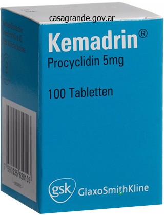
Procyclidine dosages: 5 mg
Procyclidine packs: 20 pills, 30 pills, 60 pills, 90 pills, 180 pills, 270 pills, 360 pills
Buy procyclidine overnight delivery
In gain-offunction mutation, the protein function is altered in a fashion that results in a change in the authentic operate of the gene. General FeaturesLocation of defective gene: It is on autosomes (autosomal inheritance) or the intercourse chromosomes (sex-linked inheritance). However, the inheritance will not be equal, and one gene may overpower the other in their coded attribute. The gene that overshadows the other known as the Inheritance pattern of dominant gene; the overshadowed gene is the recessive one. Autosomal Dominant Pattern of Inheritance General FeaturesLocation of mutant gene: It is on autosomes. Pattern of inheritance:Every affected individual has at least one affected mother or father. With full penetrance, all people show clinical signs With decreased penetrance, just some individuals present disease In nonpenetrance, individuals might not present any signs. Autosomal dominant: WithVariable expressivity (qualitatively or quantitatively) of disorder is the time period used for lowered penetrance only variable expression amongst people (even throughout the same family). Autosomal dominant:Expression in heterozygous stateMales and females equally affectedBoth sexes can transmit the dysfunction. Examples of autosomal dominant problems:Huntington diseaseNeurofibromatosisTuberous sclerosisMarfan syndromePuetz Jeghers polyposis. Autosomal recessive:Disease develops when each copies of gene are mutatedMales and females equally affected. When a person has one mutated gene and one normal gene, this heterozygous state is called as a service. Hematopoietic system Skeletal system Nervous system Endocrine systemSickle cell anemiaThalassemiasAlkaptonuriaFriedreich ataxiaCongenital adrenal hyperplasia Autosomal recessive disorder: Both parents will must have a mutant gene. Metabolic problems Immune techniques X-linked Dominant Disorders General Features They are very uncommon. Other X of both maternal or paternal origin is inactivated during early stage of embryonic growth. Inactivation of either the maternal or paternal X happens at random amongst all of the cells throughout about 16th day of embryonic life. Inactivation of the same X chromosome persists in all of the cells derived from every precursor cell. Genetic intercourse is set by: Y chromosome Barr body: Attached to inner side of nuclear membrane and represents inactivated X-chromosome. The inactive X could be seen within the interphase nucleus as a darkly staining small mass involved with the nuclear membrane. Y chromosome: Irrespective of the number of X chromosomes, the presence of a single Y determines the male intercourse. KaryotypingThe chromosomal structure of a cell or particular person is called the karyotype. The regular human karyotypes include 22 pairs of autosomal chromosomes and one pair of intercourse chromosomes. This contains both the quantity and appearance (photomicrograph) of full set of chromosomes. Karyotyping requires cells to be in a state of division and arresting this cell division at the metaphase of cell cycle. The more commonly used cell for chromosomal examine is circulating lymphocyte obtained from the blood sample cultured in a media. Staining: There are many staining methods utilizing specific dyes to establish particular person chromosomes. Classification of Chromosomes in Karyotyping There are numerous techniques used for examine the morphology of the chromosomes. Denver system of classification: In this system, the chromosomes are grouped from A to G based on the length and position of the centromere of the chromosomes. According to this, the chromosomes are identified based on the various banding patterns. Each chromosome reveals a characteristic banding sample (light and darkish bands) which will assist to establish them. Long and brief arm of chromosome are calledSex chromosome structure respectively: q and p. Bands and sub-bands: Each area is additional subdivided into bands and sub-bands, and these are ordered numerically as nicely.
Diseases
- Trichorhinophalangeal syndrome type II
- Osteochondritis
- Hennekam syndrome
- Kuster Majewski Hammerstein syndrome
- Pseudohermaphroditism
- Hyperparathyroidism, neonatal severe primary
- Hydantoin antenatal infection
Buy procyclidine 5mg free shipping
When extreme, it causes mucosal erosion, ulceration, hemorrhage, hematemesis and melena. Curling ulcers: They develop in the proximal duodenum with severe burns or trauma. Cushing ulcers: They develop within the stomach, duodenum, and esophagus in patients with intracranial disease. Prostaglandins defend mucosa by 1) elevated secretion of bicarbonate, 2) rising vascular perfusion, 3) inhibition of acid secretion, 4) promoting synthesis of mucin. Ulcer related to intracranial injury: They are probably as a outcome of the direct stimulation of vagal nucleicauses increased secretion of gastric acid. Morphology Gross Size and shape: It ranges from shallow erosion to deep ulcers as a lot as the mucosa; measure less than 1 cm in diameter and round in shape. Microscopy Sharply demarcated, surrounded by regular mucosa Acute inflammatory response Complete therapeutic occurs after the injurious agent/ components are eliminated. Clinical Features Most critically sick sufferers in intensive care items present microscopic evidence of gastric mucosal damage. Damaging Forces these forces are able to inducing mucosal harm and consists of two gastric secretory products: 1) hydrochloric acid and 2) pepsinogen. Gastric Acidity Ghrelin, intrinsic factor, Hydrochloric acid plays primary function in digestion but in addition can injury the gastric mucosa. Parietal cell expresses receptors for many stimulants of acid secretion, together with histamine (H2), gastrin (cholecystokinin B/gastrin receptor), and acetylcholine (muscarinic, M3). Pepsinogen, which is an inactive precursor of pepsin, is synthesized and secreted by the chief cell, discovered mainly in the gastric fundus. The acid setting throughout the abdomen leads to conversion of pepsinogen to pepsin and provides the low pH (<2. Gastric mucus barrier consists of viscid mucus (forms an unstirred layer between the epithelium and the gastric lumen) and bicarbonates 378 Exam Preparatory Manual for Undergraduates-General and Systemic Pathology c. Many of the substances, which stimulate acid secretion, additionally stimulate pepsinogen launch. Defensive Forces these are a three-level barrier composed of 1) pre-epithelial, 2) epithelial, and 3) subepithelial elements. It is shaped by several elements produced by floor epithelial cells, such as a) production of mucus, b) bicarbonate secretion, c) epithelial cell ionic transporters that maintain intracellular pH and and d) intracellular tight junctions. Actions of mucus are:Mucus layer promotes formation of an "unstirred" protective layer of fluid on the mucosa. Bicarbonate secretion: Surface epithelial cells secrete bicarbonate into the mucus bicarbonate diffuses into the unstirred mucusbuffer the hydrogen ions entering from the luminal side. It ends in a pH gradient, starting from 1 or 2 on the gastric luminal surface, and reaching to a neutrality of 6 to 7 along the epithelial cell surface. Epithelial Barrier It consists of floor epithelial cells that acts via a) restitution of damaged gastric epithelial cells, b) epithelial regeneration,c) secretion of prostaglandins and d) manufacturing of mucus (see above). Restitution: It is the process of restoration of a damaged area by the gastric epithelial cells and requires steady blood flow and an alkaline pH in the surrounding environment. Secretion of prostaglandins: Gastric mucosa secrets and techniques prostaglandin which performs a main role in gastric epithelial defense/repair. Causes persistent antral gastritis with excessive acid production may progress to pangastritis resulting multifocal atrophic gastritis increased threat of gastric adenocarcinoma. Cigarette smoking: Impairs blood flow to the mucosa and healing of mucosal harm. Alcohol, radiation therapy and chemotherapy: They trigger direct damage to mucosal bicarbonate secretion. Others ulcers High-dose corticosteroids: They suppress prostaglandin synthesis and impair~65% of gastric ulcers. Peptic ulcer: Most frequent sitesDuodenal ulcer: First a half of duodenumGastric ulcer: Lesser curvature alongside the incisura angularis. Kissing ulcers: Peptic ulcers occurring at each a posterior and anterior wall of the duodenum. Duodenum: More widespread in the first portion of the duodenum (anterior or posterior wall) than within the stomach. Stomach: Lesser curvature close to the junction (transitional zone) of the body and antrum: a. Occur at margins of the gastroduodenal anastomosis/gastrojejunostomy (anastomotic ulcer).
Safe 5 mg procyclidine
The lumen is filled with thick, mucopurulent secretions and the mucosal surface is congested. Bronchial Wall Dilatation of bronchi and bronchioles and destruction of their wall as a end result of severe irritation. Dense infiltration of persistent inflammatory cells: It primarily consists of lymphocytes and plasma cells throughout the partitions of the bronchi and bronchioles. Fibrosis of the bronchial and bronchiolar wall: It occurs in the continual instances could result in partial or full obliteration of bronchiolar lumens. Lining columnar cells might present pseudostratification and squamous metaplasia within the surrounding epithelium. These embrace staphylococci, streptococci, pneumococci, enteric organisms, and (particularly in children) Haemophilus influenzae and Pseudomonas aeruginosa. In allergic bronchopulmonary aspergillosis fungal hyphae can be seen on special stains. This is as a result of, the changes in place lead to drainage of collections of pus and secretions into the bronchi. Causes Upper respiratory tract infections: Majority are attributable to viruses (common chilly, pharyngitis). Lower respiratory tract infections: It may be due to bacteria, virus, mycoplasma, and fungus. It causes the alveoli to be crammed with inflammatory exudates and usually leads to consolidation (solidification) of lung. Lowered systemic resistance of the host: It could additionally be due toChronic diseasesImmunological deficiency: Defects in innate immunity and humoral immunodeficiency Defects in cell-mediated immunity (congenital and acquired)Treatment with immunosuppressive agentsLeukopenia. Pneumonia: Impaired local protection mechanismsLoss or suppression of the cough reflex. Impaired local defenseDamage or injury to the mucociliary equipment: It could additionally be as a outcome of cigarette smoke, viral mechanism. Portal of Entry the causative agents may enter the lung parenchyma by following routes: 1. Classification relying on the anatomic distribution Lobar pneumonia Bronchopneumonia Interstitial pneumonia 2. Clinical setting during which the an infection occurs (if no pathogen can be isolated) Community-acquired acute pneumonia Community-acquired atypical pneumonia Nosocomial pneumonia or hospital-acquired pneumonia Pneumonia in immunocompromised host Health-care related pneumonia Most widespread explanation for atypical pneumonia: Mycoplasma pneumonia. Bacterial an infection of the lung parenchyma causes the alveoli to be crammed with an inflammatory exudate and produces consolidation (solidification) of the pulmonary parenchyma. Pathogenesis Various mechanisms by which lung is contaminated are as follows: Aspiration of oropharyngeal contents: Most frequent. Classification of Bacterial Pneumonia Depending on the anatomic distribution, bacterial pneumonia could be divided into lobular bronchopneumonia and lobar pneumonia. Bacterial pneumonia: From the medical standpoint, it is very essential to determine the causative agent and decide the extent of disease. Gross and microscopic futures of produce both pattern, which is decided by the susceptibility of the patient. Bronchopneumonia, anatomically reveals patchy space of consolidation 328 Exam Preparatory Manual for Undergraduates-General and Systemic Pathology Lobar pneumonia: Most frequent trigger is Streptococcus pneumonia or pneumococcus. Morphological Stages the inflammatory response is classically divided into 4 stages: congestion, pink hepatization, grey hepatization, and resolution. The firm consistency of the affected lobe of the lung resembles that of the liver, this stage has been aptly termed pink hepatization. The fibrinous exudate within the air spaces undergoes progressive liquefaction to produce granular and semifluid debris. The liquefied material is eliminated, partly by expectoration, but mainly ingested by macrophages and drained by way of lymphatics. If not removed, it could turn out to be organized by ingrowth of capillaries and fibroblasts into the exudate. Bilateral basal location because of gravitation of secretions 2Affects extremes of age (infants or old) 3.
Procyclidine 5 mg without a prescription
Bilirubin could be displaced from binding to albumin by sulfonamides, salicylates (notably aspirin), and cholangiographic distinction media. Use of these substances in jaundiced new child infants will increase the chance of occurrence of kernicterus. Hepatic Uptake, Conjugation, and Secretion of Bilirubin Hepatocytes take up bilirubin from the sinusoidal plasma and excrete it, after conjugation with glucuronic acid, across the canalicular membrane into the bile. Despite the presence of polar carboxyl groups, bilirubin is nonpolar and lipophilic. Glucuronidation disrupts hydrogen bonds and provides polar groups to yield water-soluble pigments. Since binding of bilirubin to albumin is usually reversible, a small quantity of free bilirubin is current in plasma in equilibrium with albumin-bound bilirubin. As this free bilirubin concentration decreases, extra bilirubin is released from albumin and turns into obtainable for uptake. Alternatively, the albuminilirubin complex might bind to specific hepatocyte plasma membrane receptors, and thereby bilirubin is launched to enter the cell. The entry step appears to be carrier-mediated, is saturable, is reversible, and is competitively inhibited by sulfobromophthalein, indocyanine green, cholecystographic brokers, and a variety of other medication. After it enters hepatocytes, bilirubin is transported to the smooth endoplasmic reticulum for glucuronidation certain to a protein. Two cytosolic proteins, Z protein (fatty acid-binding protein) and ligandin (Y protein), bind bilirubin. Under normal conditions, ligandin might be the principal hepatic bilirubin-binding protein and should serve the same protecting and transport capabilities intracellularly as does albumin in plasma. It may assist limit reflux of bilirubin into plasma, since its affinity for bilirubin is a minimal of 5 times larger than that of albumin. This lack of ligandin, together with low hepatic glucuronyltransferase activity, is the possible reason for transient, "physiological," nonhemolytic, neonatal jaundice. In bile, about 85% of bilirubin is within the diglucuronide form, and the rest is within the monoglucuronide form. It is probably carrier-mediated, requires energy, is saturable, and is unaffected by bile salts. These are known as photobilirubins and are shaped when bilirubin is uncovered to blue light of 40000 nm wavelength. Mono- and diglucuronides are more water-soluble and less lipophilic than bilirubin. Conversion of bilirubin to water-soluble products is obligatory for excretion of bilirubin from hepatocytes. Metabolism of Iron and Heme Chapter 27 525 cyclization of the vinyl side group of C-3. It accommodates a seven-membered ring, is stable, is polar, and is excreted without conjugation. These observations clarify the mechanism of phototherapy commonly used for treatment of neonatal hyperbilirubinemia. Bilirubin in the Intestinal Tract Most bilirubin coming into the gut in bile is within the diglucuronide type, which is very poorly absorbed within the small and large intestines. In the lower small gut and colon, micro organism take away glucuronic acid residues and reduce bilirubin to colorless urobilinogen and stercobilinogen. Urobilinogen is excreted largely within the feces, however a small fraction is absorbed from the colon, enters the portal circulation, is removed by the liver, and is secreted into the bile. Urobilinogen excretion in urine usually quantities to 1 mg per 24 hours, as opposed to the 4080 mg (6770 mol) excreted in feces. Lack of urobilinogen in the urine and feces signifies biliary obstruction; stools are whitish ("clay-colored") owing to the absence of bile pigment. Unconjugated bilirubin is named indirect-reacting bilirubin and conjugated bilirubin as direct-reacting bilirubin. Hyperbilirubinemia might end result from elevation of unconjugated or conjugated bilirubin levels. If a larger improve happens, a point of liver dysfunction probably also happens.
Blessed Thistle. Procyclidine.
- Are there any interactions with medications?
- How does Blessed Thistle work?
- What is Blessed Thistle?
- Dosing considerations for Blessed Thistle.
- Are there safety concerns?
- Diarrhea, coughs, infections, and to promote milk flow in breast-feeding mothers, boils, wounds, and other conditions.
Source: http://www.rxlist.com/script/main/art.asp?articlekey=96137
Procyclidine 5mg without a prescription
Irreversible: Neoplasm is irreversible and persist even after the inciting stimulus is withdrawn or gone. Differentiation: It refers to the extent to which the tumor cells resemble the cell of origin. Benign tumors: They have a relatively innocent microscopic and gross characteristic. Prognosis: It is very good, can be cured by surgical elimination in many of the patients and the affected person typically survives. The term most cancers is derived from the Latin word for crab, as a result of just like a crab, malignant tumors adhere to any part that they seize on, in an obstinate method. Invasion: Malignant tumors invade or infiltrate into the adjacent tissues or constructions Metastasis: Cancers spread to distant websites (metastasize), the place the malignant cells reside, develop and once more invade. Microscopic Components of Neoplasms Tumors (both benign and malignant) consist of two primary parts: 1. The nomenclature and biologic conduct of tumors are primarily based totally on the parenchymal element of tumor. Importance of stroma: It is required for development, survival and replication of tumor (through blood supply) cells. Tumor consistency is determined by quantity of stroma:Soft and fleshy: these tumors have scanty stroma. For example, some carcinoma in female breast have stony exhausting consistency (or scirrhous). Depending on the biological habits the tumors are categorized as benign and malignant. Adenoma: Benign epithelial tumor arising from glands or forming glandular structures. Examples: Adrenocortical adenoma: It shows heterogeneous mass of adrenal cortical cells rising as a strong sheet with none glands. Pedunculated poly with stalk stalk (pedunculated polyp) or could additionally be and not using a stalk (sessile polyp). Malignant Tumors They are termed as carcinoma or sarcoma depending on the parenchymal cell of origin. These tumors have little connective tissue stroma and are fleshy (Greek sar = fleshy). Examples: Fibrosarcoma, liposarcoma, osteosarcoma, chondrosarcoma, leiomyosarcoma, and rhabdomyosarcoma. Carcinomas They are malignant neoplasms arising from epithelial cell, which may be derived from any of the three germ layers Table 7. Carcinosarcoma: It is a rare malignant tumor which reveals mixtures of carcinomatous and sarcomatous parts. Example: Mixed tumor of salivary gland (pleomorphic adenoma) is derived from a single clone (either myoepithelial or ductal reserve cell) and giving rise to two parts, particularly epithelial and myoepithelial cells (refer page 367). Teratoma: Derived from totipotent cells and incorporates tissues derived from ectoderm, endoderm and mesoderm. Teratomas They are special kinds of blended tumors derived from totipotent germ cells (normally present in ovary, testis and typically abnormally present in sequestered embryonic rest in midline). These cells have the capacity to differentiate into any of the cell sorts found in the grownup body. Thus, teratoma contains recognizable mature or immature cells or tissues consultant of multiple germ cell layer and generally all three. Classification of Teratoma Benign/mature teratoma: It consists of all mature and properly differentiated tissue. Example: ovarian cystic teratoma (dermoid cyst), in which differentiation is especially alongside ectodermal strains produces a cystic tumor lined by skin with adnexal construction (hair, sebaceous glands) and tooth buildings (refer figs 17. Immature/malignant teratoma: It consists of immature or much less well-differentiated tissue. Teratoma with malignant transformation: It is the event of malignant non-germ cell tumors from one or more germ cell layer in a teratoma. Hamartoma: Benign Example: Pulmonary chondroid hamartoma consists of islands of disorganized, but appearing, non-neoplastic overgrowth of tissue. Choristoma It is an ectopic island of regular tissue-heterotopic rest (normal tissue in an irregular Q.
Generic procyclidine 5mg visa
Deficiencies of vitamins A, C, and E, which act as antioxidants (freeradical scavengers) might improve the damage brought on by oxidants. Adenoma-carcinoma Sequence the colonic adenocarcinoma might evolve from the pre-existing adenomas, referred to as adenoma-carcinoma sequence (refer page 419). Risk is low earlier than age 40 and it increases steadily to age 50, after which it doubles with each decade. Ulcerative colitis and Crohn disease: They have elevated danger of colorectal cancer. Others: Physical inactivity, weight problems (body and abdominal), smoking, alcohol extra (especially beer) and sugar consumption are a variety of the other risk components. They may be primarily divided into 1) genetic abnormalities (that activate oncogenes or inactivate tumor suppressor genes) and 2) epigenetic abnormalities (refer web page 200). Genetic Pathways Two distinct genetic pathways have been described: A) traditional adenoma-carcinoma sequence B) microsatellite instability pathway. The lack of these tumor suppressor genes may result in uncontrolled cell proliferation. Mechanism of inactivation of tumor suppressor gene:Chromosomal deletionsMethylation of a CpG-rich zone, or CpG island (a area of some genes that incessantly consists of the promoter and transcriptional begin site). It shortens with each cell division until cell senescence develops (refer pages 36 and 211). They turn out to be unstable throughout regular mobile replication, resulting in insertion or deletion of bases within these regions. However, some microsatellites unstable during regular sequences are located within the coding or promoter area of genes concerned in regulation of cellular replication, leading cell growth. Epigenetic Abnormalities (refer page 200) Definition: Epigenetics is a reversible, heritable alteration in gene expression, which happens with out mutation. Epigenetic events could enhance development along each genetic pathways talked about above. Morphology Gross Location: Colonic adenocarcinomas may be present in any location of the colon. It could also be associated with intestinal obstruction and dilatation with attenuation and flattening of the mucosal folds of colon proximal to the tumor. They present diffuse flattening and thickening of the colon, initially involving the mucosa, however later involve the entire wall of intestine. Carcinoma colon:Exophytic polypoidal mass in the best colonAnnular constricting mass within the left colon. Most of the tumors present glands of variable size and configuration separated by average quantity of stroma. The lumen of the glands is often full of inspissated eosinophilic mucus and nuclear and mobile debris ("dirty" necrosis). The invasive element of these tumors may present stromal desmoplasiacauses firm consistency. Mucinous adenocarcinomas: They secrete ample mucin and accumulate within the intestinal wall and are related to poor prognosis. Signet-ring carcinoma: It consists of signet-ring cells much like those in gastric most cancers and represent more than 50% of the tumors. Colon cancer: Mucin production worsens the prognosis since mucin aids in tumor extension. Differences between the carcinoma of colon on proper and Tumors within the cecum and other right-sided colon cancers usually current with fatigue and left aspect. Left-sided cancers may produce occult bleeding, altered bowel habits or ache and discomfort in the left decrease quadrant. Radiology: Double-contrast barium enema: It is the radiological investigation of choice, when colonoscopy is contraindicated. Staging and Prognosis Two most necessary prognostic factors are depth of invasion and the presence or absence of lymph node metastases. Invasion into the muscularis propria reduces the survival price which is decreased additional in the presence of lymph node metastases. Carcinoembryonic antigen the tumour can spread in a longitudinal, transverse or radial course; it spreads around the intestinal Direct unfold: the tumor can unfold in a transverse, longitudinal, or radial path. Spread into the serosa, peicolic fat, lymphatics and veins within the mesentery alongside the bowel wall and generally into the peritoneal cavity. Lymphatic unfold: Tumor could unfold through lymphatics into the regional lymph nodes.
Procyclidine 5 mg sale
They are much less readily carried to the lungs and even if trapped in the lungs, they fragment into brief particles and easily ingested by macrophages. Amphibole type: the main amphibole varieties are crocidolite (blue asbestos) and amosite (brown asbestos). Amphibole form consists of straight, rigid, brittle fibers that will align themselves in the airstream and remain stable in the lung. The initial damage happens at bifurcations of small airways and ducts, the place the asbestos fibers land and penetrate. Chronic exposure and deposition of asbestos fibers lead to persistent release of fibrogenic mediators. Potentially poisonous chemicals: Inhaled toxic chemical are adsorbed onto the asbestos fibers oncogenic. For example, the adsorption of carcinogens in tobacco smoke onto asbestos fibers will increase the risk of lung carcinoma in asbestos staff. Asbestos: Exposure to increasing doses are associated with the next incidence of all asbestosrelated illnesses except mesothelioma, which is simply related to amphibole exposure. Begins as interstitial fibrosis around respiratory bronchioles and alveolar ducts and extends to contain adjoining alveolar sacs and alveoli. Later phases: the interstitial fibrosis turns into extra in depth and will involve the entire lung. The fibrosis might destroy the normal architecture of the lung to produce dilated airspaces (cystic spaces) surrounded by thick fibrous partitions produces honeycombed appearance to the involved regions. Asbestos physique is a ferruginous body golden brown, beaded and having dumb-bell form Ferruginous physique: Iron protein complex coating different inorganic particles corresponding to talc, mica and glass within the lung (not asbestos fiber). Ferruginous bodies: Other inorganic particulates/fibers may also become coated with similar ironprotein complexes called as ferruginous our bodies. Fibrosis of the lung: Lung exhibits diffuse pulmonary interstitial fibrosis, with a number of asbestos bodies. Gross: Pleural plaques seem as pearly white; well-circumscribed plaques (may be over 10 cm in diameter). Usually bilateral and most frequent on the anterior and posterolateral features of the parietal pleura and over the domes of the diaphragm. Microscopy: It consists of acellular, dense, hyalinized fibrous tissue, with many slit-like spaces organized in a parallel fashion (basket-weave pattern). Lung Cancer and Mesothelioma the workers uncovered to asbestos may develop lung carcinomas and mesotheliomas (pleural and peritoneal). Concomitant cigarette smoking markedly increases the danger of lung carcinoma but not that of mesothelioma. Pleural plaques are often asymptomatic and are visualized on radiographs as Byssinosis: Interstitial lung circumscribed densities. In the previous, the time period bronchogenic carcinoma was used for main lung most cancers, to indicate the origin from the bronchi. Etiology and Pathogenesis Tobacco Smoking Major carcinogens in tobacco smoke Table eleven. Radioactive parts and other contaminants: Arsenic, nickel, cadmium, molds, and vinyl chloride. There is strong a) statistical, b) epidemiological, c) scientific and d) experimental evidence that tobacco smoking is the most important reason for cancer of lung. Smoking additionally multiplies the risk of other carcinogenic influences corresponding to asbestos and uranium. The strongest affiliation of smoking is with squamous cell and small cell carcinoma. Clinical the danger will increase with number of cigarettes smoked and is proportional to the period four. Compared to nonsmokers, the smokers have a 10 occasions and heavy people who smoke (more than 40 cigarettes per day for several years) 60 occasions extra risk of lung most cancers. Cessation of smoking for 10 years reduces danger however to not the extent in nonsmokers. However, solely 11% of heavy people who smoke develop lung most cancers, which signifies that genetic components are involved. Carcinogenesis Tumor promotion 352 Exam Preparatory Manual for Undergraduates-General and Systemic Pathology c) Clinical proof: Microscopic changes in the lining epithelium of the respiratory tract in habitual smokers additionally revealed the carcinogenic effect of smoking. There is a optimistic relation between the depth of publicity to cigarette smoke and the histological adjustments within the respiratory epithelium.
Buy generic procyclidine 5 mg line
These enzymes are situated inside the cisternae or tough endoplasmic reticulum; because the procollagen chains enter this compartment, the hydroxylations start. All three enzymes have the same cofactor requirements: ferrous iron, -ketoglutarate, molecular oxygen, and ascorbate (vitamin C). The decreasing equivalents required for the hydroxylation response are offered by the decarboxylation of equimolar quantities of -ketoglutarate to succinate and carbon dioxide: O C H2C these reactions are catalyzed by two specific enzymes: hydroxylysyl galactosyltransferase and galactosylhydroxylysyl glucosyltransferase. The substrate has to be within the nonhelical conformation, and the glycosylation ceases when the collagen propeptides fold right into a triple helix. Thus, each hydroxylation and glycosylation should happen before triple-helix formation, which is an intracellular course of. Deficiency of galactosylhydroxylysyl glucosyltransferase has been seen within the members of kindred with a dominantly inherited disease generally recognized as epidermolysis bullosa simplex. Thus, the position of ascorbate within the hydroxylation of collagen is certainly one of its main physiological roles (see also Chapter 36). Most pathological adjustments of scurvy can be attributed to failure to synthesize collagen. Scurvy sufferers have faulty blood vessels and poor intravascular help, resulting in frequent hemorrhages, defective formation of bone and tooth, and poor wound healing. Further investigation of this dysfunction might provide details about the function of glycosylation of collagen. Formation of Disulfide Linkages and Assembly of Procollagen Polypeptides into a Triple Helix the propeptide regions of procollagen polypeptides comprise cysteine residues that may form both intra- and interchain disulfide linkages. The meeting of procollagen polypeptides into a triple helix appears to have two requirements: 1. Glycosylation of Hydroxylysyl Residues the glycosylation happens as the N-terminal ends of the polypeptide chains move into cisternae at specific hydroxylysyl residues. Extracellular Post-Translational Modification Conversion of procollagen to collagen requires at least two proteases: a procollagen aminoprotease and a procollagen carboxyprotease. The former catalyzes removing of the N-terminal propeptide, and the latter catalyzes removing of the C-terminal propeptide. The two enzymes are endopeptidases, function at neutral pH, require a divalent cation corresponding to Ca21, and present a preference for the helical conformation. Formation of Collagen Fibrils from Ordered Aggregation of Collagen Molecules the collagen molecules formed by removing of the propeptides spontaneously assemble into fibrils. At this stage, the fibrils are nonetheless immature and lack tensile energy, which is acquired by cross-linking. The initial step in cross-link formation is the oxidative deamination of -amino groups in sure lysyl and hydroxylysyl residues catalyzed by lysyl oxidase. The enzyme is a copper-dependent (probably cupric) protein, and the reaction requires molecular oxygen and pyridoxal phosphate for full exercise. In cutis laxa, the pores and skin is free and inelastic and appears to be too giant for the floor it covers. Some affected individuals exhibit deficiency of lysyl oxidase, presumably restricted to skin. The animal counterpart of lysyl oxidase deficiency is seen in several variants of aneurysm-prone mice. Lysyl oxidase undergoes irreversible inactivation when exposed to sure nitriles. Ingestion of those peas ends in lathyrism, which is characterised by multiple defects in collagen- and elastin-containing tissues. In experimental animals, lathyrism is produced by administering -aminopropionitrile throughout their lively growth period. Impaired cross-linking can even occur on account of copper deficiency, since lysyl oxidase is a copper-dependent enzyme. In experimental animals, copper deprivation causes skeletal and cardiovascular abnormalities.
Buy procyclidine 5 mg low price
The clotting instances are compared with these obtained from dilutions of pooled normal plasma, generally 1 to 10, 1 to 20, 1 to 50, and 1 to one hundred. This procedure exhibits sensitivity to the concentrations of procoagulant components and protease inhibitors including heparin, and the action of activated Protein C; and it provides details about the clotting reactions not available for elapsed time clotting exams [43]. The factor nomenclature was devised to reduce the variety of synonyms for the same component which had made description of the method of coagulation unnecessarily awkward. The bleeding time is a measure of main hemostasis, and is primarily delicate to platelet depend. Coagulation Factor Deficiencies Coagulation issue deficiencies (defects) principally affect secondary hemostasis and are most commonly mirrored in abnormal ends in clotting time-based assays. Assay strategies for assessing the operate of the hemostatic system are designed to emphasize the actual features, primary or secondary, of pathways. Both cofactor proteins are present in decreased amounts, however the proteins that have been present had been totally active. The most prevalent mutations in antithrombin result in weakened binding of heparin. Higher concentrations (doses) of heparin used as a therapeutic agent would thus be indicated for such sufferers. Chemical equilibrium is the basis for dosage adjustment in sufferers with heparin-binding mutations. The decreased extent of cross-linking of fibrin supplies the biochemical explanation. Quaternary structure variations may be brought on by postprocessing defects, not within the principal protein, but in an enzyme that processes it. Expression of genetic defects is indirect and illustrates the problem of extrapolating from gene defects to phenotype defects. A common rodenticide, brodifacoum (3-(3-(40 bromobiphenyl-4-yl)-1,2,3,4-tetrahydro-1-naphthyl)-4-hydroxycoumarin), could be deadly to people in addition to many different animals. The same mechanism of action is insufficient to fully consider the results of explicit medicine; pharmacological properties which would possibly be associated the steady-state concentrations of the drug could additionally be more important. The concentration of fibrinogen was decided by a technique that weighs the fibrin after clotting. Teaching Point Fibrinogen proteolysis or fibrin monomer polymerization may be impaired as a end result of a mutation that alters the fibrinogen molecule. Her platelet rely was measured and located to be on the high end of the normal vary. The addition of plasmin to the plasma clot resulted in virtually quick dissolution (lysis) in contrast to B10 minutes for a management clot shaped from pooled regular plasma. Teaching Point Teaching Point A historical past of bleeding by the girl, dad and mom, or different relatives should be explored. This is the outcomes of the antibiotics killing the intestinal bacteria that produce vitamin K from dietary sources of phylloquinones. Vignette 3: Activated Protein C Resistance A young African American male patient is delivered to you due to a hemarthrosis sustained after twisting his knee whereas operating. Teaching Point A 30-year-old male with recurrent thromboses is referred for diagnosis. Which parts of the hemostatic system would you initially suspect as being responsible for the thrombosis and the check results If assays for the inactivation of Factor V by activated Protein C have been normal, which of the recognized elements in the anticoagulant subsystem would you ascribe his thrombosis to Because the affected person was not beneath therapy for anything, heparin could be unlikely. Vignette 6: Von Willebrand Disease the parts are all talked about in Vignette 3. Antithrombin is indicated as being measured first because of its simplicity in comparison with Protein C and Protein S measurements. What hemostatic system elements might be responsible for the bleeding however not be detectable within the two tests for which results had been obtained Continued bleeding from the cuts was noted from a pile of blood-spotted tissues collected throughout her 3-hour wait within the emergency room!
References
- Hennerici MG, Kay R, Bogousslavsky J, et al. Intravenous ancrod for acute ischaemic stroke in the European Stroke Treatment with Ancrod Trial: a randomised controlled trial. Lancet 2006;368(9550):1871-8.
- Swift TR, Ignacio OJ. Tick paralysis: Electrophysiologic studies. Neurology. 1975;25(12):1130-1133.
- Stewart GD, Van Neste L, Delvenne P, et al: Clinical utility of an epigenetic assay to detect occult prostate cancer in histopathologically negative biopsies: results of the MATLOC study, J Urol 189:1110, 2013.
- Sarzi E, Goffart S, Seere F, et al. Twinkle helicase (PEO1) gene mutation causes mitochondrial DNA depletion. Ann Neurol 2007;62:579.
- Dockhorn RJ: Diagnostic tests for allergic disease. In Korenblat PE, Wedner HJ, editors: Allergy: Theory and Practice, Philadelphia, 1984, Grune & Stratton, p 57.
- Hoke TS, Douglas IS, Klein CL, et al. Acute renal failure after bilateral nephrectomy is associated with cytokine-mediated pulmonary injury. J Am Soc Nephrol. Jan 2007;18(1):155-164.
- Okita Y, Ando M, Minatoya K, Kitamura S, Matsuo H. Multiple pseudoaneurysms of the aortic arch, right subclavian artery, and abdominal aorta in a patient with Behcet's disease. J Vasc Surg 1998;28(4):723-6.


