
Dr Robert T Duncan
- Senior Trainee in Burns
- Wythenshawe Hospital
- Manchester
Inderal dosages: 80 mg, 40 mg
Inderal packs: 60 pills, 90 pills, 120 pills, 180 pills, 270 pills, 360 pills

Discount inderal line
Conti A et al: CyberKnife multisession stereotactic radiosurgery and hypofractionated stereotactic radiotherapy for perioptic meningiomas: intermediate-term results and radiobiological concerns. Occasionally, posttraumatic or idiopathic optic nerve sheath calcification can mimic this appearance. Note typical inferomedial displacement of the globe related to tumors of the lacrimal area. A calcified ocular mass in a baby represents retinoblastoma until proven in any other case. Wyse E et al: A review of the literature for intra-arterial chemotherapy used to treat retinoblastoma. Note distinguished enhancement of every iris; anterior section enhancement is associated with extra aggressive tumor habits. Note intact skinny lines of enhancing choroid and hypointense sclera, indicating absence of invasion of those constructions. There is subtle thickening and enhancement of the best optic nerve, indicating postlaminar invasion. The left postlaminar optic nerve demonstrates uneven enlargement and enhancement. There is a cumbersome enhancing suprasellar and parasellar mass, with extension along dural reflections. Ocular melanomas distant from the macula may be quite giant earlier than vital visible defects are obvious. Lenticular-shaped delicate hyperintensity lateral to the enhancing lesion is compatible with associated retinal detachment. The mushroom-shaped configuration with waist-like narrowing at the tumor base indicates that invasion of Bruch membrane is probably going. Bulky extraocular tumor is seen extending posteriorly by way of the optic nerve head. The very large size of this melanoma and the presence of extraocular extension are each poor prognostic components. This is a variant case, each due to the massive quantity of extraocular disease and its extremely infiltrative appearance. There are bilateral lacrimal gland lesions along with intraconal, conal, and extraconal involvement. The superolateral bony orbit is invaded by this lacrimal gland carcinoma, and the globe is displaced inferomedially. The mass is closely apposed to the globe with scleral irregularity suspicious for invasion. T1 precontrast photographs show lesion margins against the contrast of skull base marrow fats. T1 additionally reveals highsignal subacute blood and intralesion high-velocity flow voids to finest benefit. Enhanced, fat-saturated T1 sequences outline enhancement traits of the lesion in question. The prenasal space is a transient prenatal region separating nasal bones and cartilaginous nasal capsule. The anterior neuropore extends from intracranial area to prenasal area and briefly contacts skin at the bridge of the nostril but involutes previous to start. The prenasal area reduces to a small canal anterior to the crista galli called the foramen cecum. Melanoma, N Lacrimal gland carcinoma, O Central cranium base Sella: Pituitary macroadenoma Clivus: Chordoma, ecchordosis physaliphora Petrooccipital fissure: Chondrosarcoma Meckel cave: Trigeminal schwannoma T-bone: Tumor Endolymphatic sac tumor T-bone: Tumor-like lesions Acquired cholesteatoma Congenital cholesteatoma Cholesterol granuloma Posterior cranium base Clivus (occipital bone): Chordoma Jugular foramen Glomus jugulare paraganglioma Jugular foramen schwannoma Jugular foramen meningioma Hypoglossal canal: Hypoglossal schwannoma diameter is ~ four mm. Ossification of the crista galli and cribriform plate begins at 2 months and is type of full by 24 months. Persistence of the craniopharyngeal canal (remnant of Rathke pouch) could occur between the presphenoid and basisphenoid. Persistence of the median basal canal may be seen between the basioccipital ossification facilities. Nasopharyngeal carcinoma instantly accesses the intracranial compartment via the foramen lacerum (perivascular spread). Borges A: Skull base tumours half I: imaging method, anatomy and anterior cranium base tumours.
Discount 10 mg inderal
It primarily happens on sun-exposed pores and skin, in general in any hair-bearing space, however may happen at sites with restricted or no sun exposure. Aggregates of basaloid tumor cells in the superficial dermis are linked to the dermis. Typically a quantity of such foci are current, separated from one another by "skip" areas of normal skin (in two-dimensional view). B, At the periphery of the tumor nodule, basaloid cells type a picket fence or "palisading" arrangement. Such basaloid nests are usually multifocal and separated from one another by intervening areas of regular dermis (also known as skip areas). We have just lately proven that measurement of the depth of invasion (similar to Breslow thickness) helps predict response to topical remedy. Thin cords and oddly formed aggregates of tumor cells are dispersed in a haphazard infiltrative sample. Thin anastomosing cords of basaloid cells are present in affiliation with a fibrous stroma. A, A small nodule of cytologically bland basaloid cells is present with small keratocysts. On event, one also finds dendritic melanocytes within the tumor compartment (melanocytes and Langerhans cells). Squamous differentiation may happen in deeper infiltrative elements of the tumor, particularly in recurrent tumors. Basal cell carcinomas could collide with a selection of epithelial and nonepithelial tumors. However, problems can arise from histologic overlap with other basaloid epithelial neoplasms and from inadequate histologic sampling. Biphenotypical carcinomas often have a predominant basal cell appearance on the preliminary biopsy, however with subsequent recurrences, the squamous part tends to dominate and dictate the clinical conduct. For early superficial lesions, local destructive modalities, similar to cryotherapy, may be enough. For facial lesions, surgery with frozen section examination is the remedy of alternative. Postoperative or intraoperative full circumferential peripheral and deep margin evaluation with frozen or permanent sections is important for large and infiltrative lesions. Identification of true tubule formation and attention to the overall progress pattern or immunohistochemistry are useful. Sweat gland tumors, together with adenoid cystic carcinomas, are inclined to be optimistic for the latter three markers. The tumors are composed of two main cell types: (1) basaloid cells with matrical options are organized in lobules and sheets or cords and bands and (2) "ghost" or "shadow" cells as seen in pilomatricoma. Pilomatrix carcinoma differs from pilomatricoma primarily by its infiltrative growth pattern and the presence of cytologic atypia. Basal cell carcinoma, which can show focal matrical differentiation, differs from pilomatrix carcinoma (and pilomatricoma) by the presence of typical features of Pilomatrix carcinoma. Not enough bona fide instances have been reported to permit a clear description of related clinical and histologic features. Trichilemmal carcinomas are described in the literature as infiltrative dermal tumors with outer root sheath differentiation, sometimes exhibiting a clear, glycogen-rich cytoplasm. They are usually cured by excision, but uncommon tumors metastasize to lymph nodes and visceral websites. Additional options suggestive of trichilemmal carcinoma include peripheral cell palisading and trichilemmal keratinization. They may show tubular, cribriform, papillary, micropapillary, or blended progress patterns. The ductal lining is bilayered, with luminal cuboidal secretory cells and peripheral cuboidal or flat myoepithelial cells.
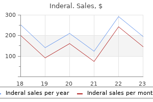
Generic 10 mg inderal otc
Leg veins less than 1 mm in diameter are additionally successfully handled with pulsed dye lasers using a pulse period of as much as 20 msec. One of the newer pulsed dye lasers even has a handpiece to treat pigmented lesions. This is a compression handpiece designed to bodily compress out the blood, thereby eradicating one of many competing chromophores of this wavelength. Theoretically, gentle heating of the superficial dermal layer will stimulate the production of recent collagen. Pulsed dye lasers at low to moderate fluences (nonpurpuric settings) are used for this nonablative resurfacing. Omi T, Kawana S, Sato S, et al: Cutaneous immunological activation elicited by a low-fluence pulsed dye laser, Br J Dermatol 153(Suppl 1):57�62, 2005. There could also be significant post-treatment purpura that takes 7 to 10 days to resolve. This purpura outcomes from the explosive optical-acoustic pulse generated by the pulsed dye lasers. The newer pulsed dye lasers are capable of treat lesions with out purpura by utilizing longer pulse durations in the vary of 10 msec (Table 53-5). The standard wavelength of light emitted is 1064 nm, which is in the near-infrared spectrum. This laser has also been used for hair elimination in tanned patients, but I would advocate avoiding laser treatments with just lately tanned pores and skin. These long-pulsed lasers are also very helpful for treating vascular lesions together with leg veins and larger vessels on the nose. The primary benefit over pulsed dye lasers is the dearth of purpura after treatment. This laser has also been helpful in treating dermal pigmented lesions corresponding to nevus of Ota and nevus of Ito. The most sensational feature about this stone, however, is its surprising capacity to change its colour. Green or bluish-green in daylight, alexandrite turns a gentle shade of pink, purplish-red, or raspberry purple in incandescent mild. An alexandrite laser is a stable state laser in which chromium ions (Cr-3), on the quantity of 0. The Q-switched alexandrite laser is efficient in eradicating green and black tattoo pigment. The long-pulsed laser has been reported to successfully remove leg veins that are 1 to 2 mm in diameter and blue in colour. This laser has lately also been reported to effectively deal with brown lentigines on the face and hands (Table 53-7). The ruby laser has an lively medium of aluminum oxide (Al2O3) that has been chromium-doped meaning that some of the aluminum (Al+3) atoms have been replaced with chromium (Cr+3) atoms. The Q-switched ruby laser mild is nicely absorbed by black, blue, and green tattoo pigment. The Q-switched ruby lasers have additionally been utilized in treating some dermal pigmentation abnormalities corresponding to nevus of Ota. The long-pulsed ruby laser has been used primarily for laser hair elimination (Table 53-8). Diode lasers use microscopic chips of gallium arsenide or other semiconductors to generate coherent gentle. The vitality of the sunshine is generated by the variations between vitality levels inside the semiconductor. The benefit of the diode lasers is lower value, a variety of wavelengths, and smaller lasers. The 810-nm diode lasers are primarily used for hair removing and, occasionally, for treatment of vascular lesions. The 1320-nm diode lasers have been used for nonablative laser resurfacing and treatment of acne scarring. The 1450-nm diode lasers have been used for nonablative laser resurfacing, therapy of zits scarring, and the therapy of active acne (Table 53-9). The concept is that melanin and elastic tissue might be removed, and the thermal damage will result in collagen stimulation. Since this first laser, there have been many nonablative lasers with a big selection of wavelengths and theoretical mechanisms.

Cheap inderal
Bullous allergic contact dermatitis in a florist because of slicing and handling carnations. Positive patch take a look at reactions to the rubber accelerators thiuram and carbamates present in rubber gloves in a health care worker. Studies have shown that delay of analysis for more than 1 12 months and continual publicity are essential components for chronicity. Belsito D: Occupational contact dermatitis: etiology, prevalence, and resultant impairment /disability, J Am Acad Dermatol 53:303�313, 2005. Allergic contact dermatitis happens commonly with rubber gloves containing the chemical substances thiuram, mercaptobenzothiazole, and carbamates, that are "rubber accelerator" chemicals used to pace up the vulcanization process. Some allergens can penetrate varied glove materials and turn out to be trapped towards the skin. Patients should be instructed to avoid both extreme water publicity and frequent hand washing. Hands could be shielded from the weather by utilizing cotton glove liners to absorb perspiration inside a correct protecting glove for the job. Moisturizers should be used immediately after wetting the palms or every time they appear dry and scaling. Topical corticosteroids are the mainstay of remedy for occupational contact dermatitis, with systemic steroids reserved for acute, extreme situations. With remedy and hand protection, many staff can and do continue to work regardless of a hand dermatitis. Most employees suffer monetary and social consequences from altering occupations and do finest with environmental modifications that permit them to stay on their job. The basic aim of prevention is to establish dangers to the pores and skin and discover ways to remove publicity or a minimum of to minimize it. For example, allergenic biocides in chopping oils could be changed by different preservatives. For instance, screens or filters can be used to stop chemical splashes or addition of hand instruments can be used to grasp irritating compounds. Prompt and proper hand washing and use of frequent moisturizers must be inspired. Personal protecting tools, including aprons, boots, visors, sleeves, and gloves, must take into account the specific needs of every state of affairs and publicity. Many studies confirm that a high percentage of dermatology sufferers have coexistent psychiatric morbidity. We do know that the pores and skin and nervous system have the identical embryologic origin, and that tactile stimulation is critical for full neuropsychological growth, but many questions remain. Recent biomedical advances have begun to take psychocutaneous illness out of the realm of speculation and myth. As we examine shared symptom complexes and responses to pharmacologic intervention, many psychocutaneous problems can now be considered in terms of neurotransmitters and their receptors, with all of the inherent implications for treatment. Instead, our deeper understanding of psychocutaneous illness can only lead to improved patient care. Psychocutaneous illness can be categorized into three major classes: � Primary psychiatric disorders with dermatologic manifestations � Primary dermatologic disorders that lead to secondary psychiatric problems. This figure is troublesome to assess; nonetheless, some studies have reported that more than 40% of all dermatologic sufferers have associated psychological morbidity. These patients falsely imagine that their skin is infested with parasites and this is frequently related to cutaneous dysesthesia. They typically describe insects mating, laying eggs, and crawling round of their pores and skin. They develop elaborate purification rituals and are often well known to pest management organizations. Of notice is the reality that the delusion is commonly referred to as a monosymptomatic hypochondriacal psychosis. Scale, scabs, and hair introduced in by a affected person with delusions of parasitosis who insists that these are parasites. One should rule out all different possible reasons that a patient would possibly complain that she or he is infested with parasites. Delusions, like hallucinations, are psychotic symptoms which would possibly be theorized to end result from increased ranges of dopamine in elements of the brain. Patients with delusions of parasitosis have been shown to respond to neuroleptics; particularly, the dopamine antagonist pimozide (Orap) is, historically, the commonly used treatment.

Purchase cheap inderal
Thus, if a pores and skin eruption is photodistributed, even with no definite history of exacerbation following solar publicity, many dermatologists classify it as a photosensitive dermatosis. A photodistributed eruption impacts the pores and skin in a characteristic distribution, affecting the convex surfaces of face, exterior areas of forearms and arms, dorsal hands, V-area of higher chest, lateral sides and posterior of the neck, and any other area uncovered to the sun. It characteristically spares the ocular/eyelid space, beneath the nose and lower lip, inframandibular chin/neck, and internal parts of arms and forearms, and clothing-covered websites. In photocontact dermatitis, the affected areas are these areas with publicity to both daylight and the causative topical agent. It could happen within minutes to hours of publicity, though it might also be delayed for a day or two. Photoallergic reactions typically happen 1 to 3 days after exposure (with the exception of photo voltaic urticaria, which is immediate). Photoallergic reactions are also photodistributed, however often have extension of the cutaneous reaction onto coated areas or even distant sites in an autoeczematous type of eruption. Sunburn-like erythema on the cheeks, neck, V-area of the chest, and dorsal forearms. A, Photoallergic drug eruption as a outcome of oral compazine demonstrating marked erythema and swelling of the dorsum of the hands, arms, and V of the chest. Numerous merchandise (soaps, perfumes, sunscreens) could produce photoallergic contact dermatitis in some people. Numerous medicine, both prescription and nonprescription, can often produce photosensitive reactions. Pruritus is a typical complaint related to sure diseases (such as photoallergic contact dermatitis), whereas pain or burning is extra generally related to phototoxic problems. In persistent mild reactivity, photodermatitis believed to be triggered by topical or systemic medication persists lengthy after the presumed causative agent has been discontinued. Elderly man with continual, extremely pruritic photosensitivity with erythema, scale, pigmentary changes, and lichenification of the pores and skin. Erythematous, scaly plaque on the lateral neck, which tended to recur each spring. Some consider that the first occasion in these circumstances is a photocontact dermatitis (photoallergic dermatitis) which persists as a result of persistent low-grade exposure, and in rare circumstances, even progressing to an "antigendriven" type of mycosis fungoides, though that is controversial. Phototoxic reactions clinically and symptomatically resemble sunburn while photoallergic reactions resemble dermatitis. Patients characteristically report the onset of skin illness beginning with solar publicity in spring or early summer season. Patients typically show gradual enchancment with continuing sun exposure, a phenomenon termed "hardening. Lesions are photodistributed, often on the face and neck, chest, and dorsal arms and hands. Native American baby with pruritic photosensitive dermatitis of nostril, cheeks, and chin. Thus, you will need to exclude different causes of photosensitive dermatoses, corresponding to lupus erythematosus, porphyrias, and photoallergic eruptions from systemic or topical medications. In this population, the family history is usually optimistic, though sporadic cases do happen. Ross G, Foley P, Baker C: Actinic prurigo, Photodermal Photoimmunol Photomed 24:272�275, 2008. The urticarial papules or plaques often develop on uncovered areas within minutes of solar exposure and are accompanied by pain or pruritus. Solar urticaria is a photoallergic response (type I hypersensitivity response) and is most probably IgE mediated, though direct degranulation of mast cells may also be a pathogenic mechanism. Erythropoietic protoporphyria could current equally and must be dominated out with porphyrin research. Du-Thanh A, Debu A, Lalheve P, et al: Solar urticaria: a time-extended retrospective collection of 61 patients and evaluate of the literature, Eur J Dermatol 23:202�207, 2013. In the neonatal period, photodermatoses are uncommon, probably due in part to the minimal degree of exposure to daylight.
Order inderal amex
The texture of the scalp may remain gentle and supple, although generally induration or firmness is palpable. People who turn out to be bald have hair follicles which are genetically programmed to miniaturize beneath the influence of postpubertal androgens. Probably, a quantity of genes (inherited from each mom and father) affect the severity of balding. Except in very marked and long-standing balding, very nice, short hairs can be seen exiting from follicular orifices if a magnifying lens is used. About one third of balding sufferers who use topical minoxidil solution expertise important (cosmetically obvious) hair regrowth. Oral finasteride, a 5-reductase inhibitor, is somewhat simpler, and can be used in combination with topical minoxidil. In the absence of elevated circulating androgens, nonspecific therapy directed at suppressing ovarian androgen production or blocking the peripheral effect of androgens is typically tried. Oral contraceptive brokers (to suppress ovarian androgen production) and spironolactone are most often utilized for this purpose. Topical minoxidil answer can additionally be useful, however oral finasteride is seldom utilized in women. Men, and infrequently girls, can achieve everlasting cosmetic enchancment by present process a hair transplantation procedure. Hair follicles from the occipital area (donor site) are moved to the balding space (recipient site). The procedure is tedious and expensive, however the beauty outcomes could be fairly good. Central, centrifugal, cicatricial alopecia, a common form of hair loss in the African-American inhabitants. In this patient, the smooth skin, devoid of most follicular openings, displays light like a mirror. A large bald patch studded with small inflammatory papules surrounded by smaller, similar lesions. B, A patch of alopecia areata showing shorter, thinner, and fewer deeply pigmented hairs rising throughout the bald zone. Although many forms of alopecia can lead to a circular bald patch, the most common causes are tinea capitis and alopecia areata. Tinea capitis is a superficial fungal infection with a predilection for children, particularly black children. The surface of the pores and skin is scaly and generally infected, and small darkish stubs of hair ("black dots") could additionally be scattered inside the affected area. In this condition, the hair shaft is invaded and replaced by myriad circular fungal spores. Alopecia areata also commonly affects children, however adults more typically develop the condition. In alopecia areata, the affected areas may be totally hairless, however the scalp floor appears in any other case normal, without scaling and minimal, if any, erythema. When a solitary lesion of alopecia areata is small (<5 cm in diameter), no treatment could also be wanted. The prognosis for such a lesion is great, and spontaneous regrowth usually occurs. Intralesional corticosteroids, and typically potent topical corticosteroids, may hasten regrowth. The use of systemic corticosteroids to treat in depth alopecia areata is controversial. Although spontaneous regrowth may happen even in alopecia totalis, no remedy has been discovered to be persistently safe and effective for extreme disease. For now, topical immunotherapy with chemical substances causing allergic contact dermatitis. Systemic antifungal brokers are required to eradicate the spores that invade affected hair shafts. Ultra-microsized formulations of griseofulvin may be given in half the dose required with microsized types of the drug. The typical patient is an adolescent lady, though the condition can have an effect on youngsters of both sexes as well as adults.
Diseases
- Deciduous skin
- LyP (lymphomatoid papulosis)
- Pycnodysostosis
- Microgastria short stature diabetes
- Eosinophilic granuloma
- Microcephalic osteodysplastic primordial dwarfism
- Ackerman syndrome
- Metaphyseal dysplasia Pyle type
- Carotid artery dissection
Buy 80mg inderal with amex
Nicolin G et al: Natural historical past and end result of optic pathway gliomas in children. Schupper A et al: Optic-pathway glioma: natural historical past demonstrated by a brand new empirical rating. Taylor T et al: Radiological classification of optic pathway gliomas: experience of a modified practical classification system. Thiagalingam S et al: Neurofibromatosis kind 1 and optic pathway gliomas: follow-up of fifty four sufferers. Laithier V et al: Progression-free survival in kids with optic pathway tumors: dependence on age and the standard of the response to chemotherapy-results of the primary French potential research for the French Society of Pediatric Oncology. A peripheral zone of higher sign depth is appropriate with associated arachnoid hyperplasia. Between the ossification centers of presphenoid is a cartilaginous gap referred to as the olivary eminence, which is obliterated shortly after start. In the midline, observe the craniopharyngeal canal, sphenooccipital synchondrosis, and median basal canal. The sphenooccipital synchondrosis fuses over the primary 20 years of life whereas the craniopharyngeal and median basal canals are rarely persistent into childhood. Four areas have key interactions with skull base: Masticator, parotid, carotid, and pharyngeal mucosal spaces. The pharyngeal mucosal area abuts the foramen lacerum, which is covered by fibrocartilage in life. Neural constructions have been eliminated on proper, permitting visualization of numerous perforations within the cribriform plate, via which afferent fibers from olfactory mucosa pass to type the olfactory bulb. The posterior margin of the anterior skull base is fashioned by the lesser wing of sphenoid and planum sphenoidale. Note the foramen cecum, a small pit anterior to the crista galli, bounded anteriorly by frontal bone and posteriorly by ethmoid bone. If the anterior neuropore persists, an enlarged foramen cecum, bifid crista galli, and epidermoid along the neuropore tract are attainable. A regular stalk of dura extends via the foramen cecum to pores and skin (anterior neuropore). The greater wing of the sphenoid bone varieties the anterior wall of the middle cranial fossa. The posterior limit of the central skull base is the dorsum sella medially and the petrous ridge laterally. The neural constructions are proven on the left while the bony landmarks are seen on the best. The anterior boundary of posterior cranium base is clivus medially and petrous ridge laterally. The major foramina are the foramen magnum, porus acusticus, jugular foramen, and hypoglossal canal. Notice that the jugular foramen connects anteriorly with the petrooccipital fissure. The midbrain and pons in addition to the left half of the tentorium cerebelli have been eliminated. Notice the transverse sinus is within the wall of the occipital bone while the sigmoid sinus is in the medial wall of the temporal bone. Changes in cell biology, extracellular matrix components, angiogenesis and genetics. Notice proper posterolateral displacement of the basilar artery & compression of the pons without parenchymal edema from this slow-growing tumor. One hundred sufferers irradiated by a 3D conformal approach combining photon and proton beams. Chordoma Skull Base Lesions (Left) Axial graphic illustrates a large clival chordoma pushing posteriorly to indent the low pons & basilar artery.
Generic inderal 40 mg on line
This youngster has continual lead toxicity attributable to absorption of the lead-based pigments associated to her surmacosmetic plumbism. Surma usually has lead-based pigments and has created problems with lead toxicity in several Asian communities in the United Kingdom. In the Middle East, a similar conventional eye cosmetic has additionally been demonstrated to incessantly comprise high levels of lead. Moxa is from mokusa, the Japanese word for wormwood (Artemisia moxa of the sagebrush and absinthe genus), a commonly used combustible medicinal herb. Moxibustion is the ancient oriental medical practice of igniting medicinal herbs on the pores and skin. The sites on which moxibustion are performed are sometimes the identical as those utilized in acupuncture, and it, together with cupping and acupressure, is considered to be a nonneedle form of acupuncture. Bloodletting, which has widespread roots in plenty of historical traditions of drugs, is a legitimate therapy of a handful of medical conditions, including hemochromatosis and porphyria cutanea tarda (although this process is now referred to as "therapeutic phlebotomy"). In earlier centuries, bloodletting was thought of to be a therapy possibility for all kinds of medical conditions and the remedy of choice for pneumonia. While the apply has persisted longer in certain areas of the world, its efficacy in all however a limited number of circumstances, such as these listed above, has been disproven. Papavramidou N, Thomaidis V, Fiska A: the traditional surgical bloodletting technique of arteriotomy, J Vasc Surg fifty four:1842�1844, 2011. In a big survey of more than 6300 acupuncture patients within the United Kingdom, sufferers reported at least one antagonistic event in about 10% of cases, the commonest of which was bruising and bleeding. There have been reports of ample petechiae (in one case, resembling meningococcemia) attributable to acupuncture needles. Pyoderma, extended anesthesia, needle breakage, burns, itching, argyria, ulcers, postinflammatory hyperpigmentation, overseas physique granuloma, "carcinoma of the pores and skin," and psoriatic Koebner phenomenon have also been reported. Yamashita H, Tsukayama H, Taanno Y, et al: Adverse events in acupuncture and moxibustion remedy: a six-year survey at a national clinic in Japan, J Altern Complement Med 5:229�236, 1999. It is advocated to be helpful in the remedy of a broad range of illnesses similar to respiratory illnesses. Suction is created by a variety of totally different strategies including mechanical gadgets. Archaeological evidence, such as human stays, reveals that tattooing was part of indigenous cultures worldwide. For no matter causes, tattoos had been used in historic Europe, the Mediterranean area and Middle East, southern Asia, northern Japan, the Americas, and throughout the Pacific islands. Levy J, Sewell M, Goldstein N: A short history of tattooing, J Dermatol Surg Oncol 5:851�856, 1979. Chinese girl with quite a few sharply demarcated hemorrhagic lesions of the again as a end result of cupping remedy. The traditional patterns of Polynesian tattoos are distinctive for every island or island group. The Marquesan Islanders of French Polynesia as quickly as utilized tattoos to almost the complete body. The apply is experiencing a cultural resurgence in many Polynesian groups right now. Japanese tattoos (horimono) are often regarded as probably the most skillful and artistically ready. European sailors adopted the habit throughout voyages to the Polynesian islands in the 18th century. Many ethnic groups, cultures, and even subcultures, together with sailors, have tattoos with particular meanings. Other examples of tattoos with specific meanings embrace an anchor, meaning that the sailor had sailed the Atlantic Ocean, or a completely rigged sail ship, indicating that the sailor had sailed round Cape Horn. The Ice Man is the name given to the 5200-year-old frozen corpse of a Bronze Age hunter discovered preserved within the ice of a Tyrolean glacier on the border of Austria and Italy. Ornamentation and beautification, group identification, safety (from sickness or evil), and therapy. It symbolizes the ceremony of passage by initiates coming into the predominantly however not solely black school fraternity Omega Psi Phi. Kani is the Fijian word for kava dermopathy, which is an acquired ichthyosiform disorder attributable to extreme consumption of kava (called yaqona in Fiji).
Order inderal now
Also, in distinction to hypertrophic scars, keloids uncommonly bear involution, and so they regularly proliferate nicely beyond the bounds of the unique trauma. Some keloids, particularly on the sternum or higher back, even appear to develop without identifiable preceding trauma. A, Multiple, small, typical exophytic, light brown acrochordons of the axilla associated with acanthosis nigricans. In early lesions, it might be impossible to make this distinction, but in developed lesions, the options listed in Table 42-2 are helpful. Many factors can predispose individuals to develop hypertrophic scars and keloids: � Certain drugs. Thermal burns, with their associated extreme tissue damage, generally produce hypertrophic scars or keloids. Blacks are 2 to 19 instances and Asians 3 to 5 times extra likely than Caucasians to develop keloids. The tendency for a number of keloids to occur in households additionally suggests a genetic predisposition. Lacombe D, Morice-Picard F: Rare genetic ailments, signalling pathways, and keloid scar formation, Br J Dermatol 171:452�453, 2014. Typical acquired digital fibrokeratoma demonstrating an exophytic firm papule with a hyperkeratotic collarette. In general, hypertrophic scars respond well to much less aggressive therapy, corresponding to potent topical steroids, intralesional steroids, chronic strain dressings, and even persistent occlusive dressings with medical Silastic gel sheeting. Keloids demand more aggressive remedy with long-term precautions to prevent recurrence. Surgical excision, corticosteroid injection, and cryotherapy alone can be used however are sometimes not effective. A latest research has advised that incomplete excision of keloids is associated with an elevated likelihood of recurrence. Radiation has been employed with good results however must be used with warning and solely by an experienced doctor. Surgical excision with subsequent steroid injection is probably the simplest approach. One routine requires steroid injection to the surgical website 1 to four weeks postoperatively after which monthly for 6 months. These solitary, firm, hyperkeratotic papules most commonly occur round interphalangeal joints but could occur on any website of the hands or toes. Histologically, they show overlying hyperkeratosis and thickened collagen bundles. Some authorities consider that these lesions come up as a reaction to recurring trauma. Nodular fasciitis (pseudocarcinomatous nodular fasciitis) is an unusual, benign, myofibroblastic tumor that many consider to be a pseudoneoplastic or reactive development. It may happen at any age however mostly presents in younger adults as a solitary, quickly rising, subcutaneous nodule on the extremities. The nodule could additionally be painful, is typically 1 to 3 cm in measurement, and will adhere to the underlying fascia. Simple excision is healing, but untreated lesions have been documented to regress spontaneously. The importance of this lesion is that, histologically, it may be confused with malignant fibrous neoplasms, as a result of the fibroblasts are massive and pleomorphic and mitotic figures are comparatively widespread. Connective tissue nevus is a term used for cutaneous hamartomas composed primarily of collagen (collagenomas), elastin (elastomas), or a combination of those two. While these are produced by fibroblasts, connective tissue nevi are typically no more cellular than regular dermis. Connective tissue nevi composed primarily of collagen are skin-colored, while these composed primarily of elastin could also be skin-colored or yellowish. However, some of these nevi function cutaneous markers for other systemic syndromes. Collagenomas called shagreen patches are regularly seen in patients with tuberous sclerosis. Multiple connective tissue nevi referred to as dermatofibrosis lenticularis disseminata are composed of an admixture of elastic fibers and collagen and are the cutaneous marker for Buschke-Ollendorff syndrome.
Proven inderal 40 mg
More systematic research of those entities with regard to the utility of staining refined cases with p53 and p16 are wanted. The calcified corneocytes can happen within the epithelium or in the cornified portions of the tumor. A, Crateriform squamous proliferation with central keratin plug and lobular development. B, Keratinocytes with bland nuclei, abundant pale eosinophilic cytoplasm, and individually necrotic keratinocytes. C, Another cub-shaped squamous proliferation with in depth hyperkeratosis from the identical patient. D, Downward lobular bulbous epithelial progress of well-differentiated keratinocytes with bland nuclear options extending to bone. A historical past of trauma is widespread, and there could additionally be underlying bony abnormalities as properly, similar to exostoses. They consist of eosinophilic keratinocytes and will form small cysts that may mimic ductal epithelium, forming an internal zone of keratinization. Whether these inclusions display real adnexal differentiation is a matter for additional study. A single giant solitary cystic inclusion, typically painful and associated with parrot-beak and clubbing deformity of the nail, and osteolysis, is referred to as an epidermoid implantation cyst. Multiple inclusions may be deemed type 1 or 2 depending on their extent, with sort 1 being associated with limited clinical findings and superficial histology and type 2 with extra notable scientific adjustments and extra extensive aggregates histologically. As is the case for different nail tumors, they often have a deceptive clinical appearance resembling a dermatitis, paronychia, fungal infection, or nonspecific ulcer with adjoining nail dystrophy. It typically presents as a nodule near the proximal nail fold, with associated yellow thickening of the nail plate (also known as xanthopachyonychia), transverse overcurvature of the nail plate, and proximal splinter hemorrhages. If eliminated intact, it could be noticed to comprise both stromal and epithelial parts of the matrix and possibly nail bed. There are additionally downward and inward projections of matrical epithelium forming deep channels interdigitating with epithelium ("glove-finger" appearance), and a fibrocellular stroma of variable cellularity, once in a while considerably myxoid. These could replicate the same phenomenon as comparable cells in the benign pleomorphic fibroma, for example. The nail plate past the lunula is thick, with cavities containing loculated serum, and infrequently sometimes hemorrhage. Histologically, the authors describe three variants: acanthotic, papillomatous, and keratogenous. B, There is reduplicated nail matrix epithelium, with the superficial features of the keratogenous zone apposed to the proximal nail plate. C, the proximal fibroepithelial portion consists of a fibrous stroma of various cellularity. They all current as skin-toned papules, usually with overlying hyperkeratosis, usually in periungual areas, typically resting on the nail plate. Single or multiple lesions may be present, the latter particularly in the setting of tuberous sclerosis. It is a slow-growing, painless nodule that has a predilection for the arms and feet, and involvement of the nail unit happens in 50% of the instances. It may mimic an acral angiofibroma, and in one case report, occupied the whole nail unit. Especially if oriented transversely as on this case, the big loculations containing serum and often hemorrhage are pretty distinctive. It is important to be familiar with this benign neoplasm because the differential diagnosis contains many benign and some malignant myxoid neoplasms, together with myxoid neurofibroma, sclerosing perineurioma, superficial angiomyxoma, myxoid derma- tofibrosarcoma protuberans, low-grade myxofibrosarcoma, and myxoinflammatory fibroblastic sarcoma. Typically, it accommodates sheets of histiocytes, usually foamy, and sometimes with multinucleate osteoclast-like giant cells, hemorrhage, hemosiderin, siderophages, and typically a sclerotic stroma. A, the tumor consists of an acanthotic matrical epithelium, forming small whorls of incipient nail plate. B and C, the tumor forms so-called prekeratogenous and keratogenous areas, recapitulating normal nail plate formation by the matrix.
References
- Childs C, Wieloch T, Lecky F, Machin G, Harris B, Stocchetti N. Report of a consensus meeting on human brain temperature after severe traumatic brain injury: its measurement and management during pyrexia. Front Neurol. 2010;1:146.
- Nathoe HM, van Dijk D, Jansen EW, et al: A comparison of on-pump and off-pump coronary bypass surgery in low-risk patients, N Engl J Med 348(5):394-402, 2003.
- Macdonald JS, Smalley SR, Benedetti J, et al. Chemoradiotherapy after surgery compared with surgery alone for adenocarcinoma of the stomach or gastroesophageal junction. N Engl J Med 2001;345(10):725-730.
- Wang C, Li L, Diao M, et al: Single-incision laparoscopic-assisted anorectoplasty for the management of persistent cloaca, J Laparoendosc Adv Surg Tech A 26(4):328n333, 2016.
- Rowe RD, Freedom RM, Mehrizi A, Bloom KR. The neonate with congenital heart disease. Major Problems in clinical pediatrics. 2nd edition. W.B. Saunders, Philadelphia. 1981;5:101-9, 515-28.
- Cui Y, Li G, Li S, et al. Designs for linkage analysis and association studies of complex diseases. Methods Mol Biol 2010; 620: 219-242.
- Fisher CM. Pathological observations in hypertensive cerebral hemorrhage. J Neuropathol Exp Neurol 1971;30:536.
- Mumtaz H, Williams V, Hauer-Jensen M, et al: Central venous catheter placement in patients with disorders of hemostasis. Am J Surg 180:503, 2000.