
Helen W. Boucher, M.D., F.A.C.P.
- Assistant Professor of Medicine
- Tufts University School of Medicine
- Director
- Fellowship Program
- Division of Geographic Medicine and
- Infectious Diseases
- Tufts Medical Center
- Boston, Massachusetts
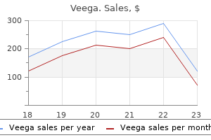
Veega dosages: 100 mg, 75 mg, 50 mg, 25 mg
Veega packs: 10 pills, 20 pills, 30 pills, 60 pills, 90 pills, 120 pills, 180 pills, 270 pills, 360 pills

Buy line veega
First branchial cleft anomalies are the results of branchial cleft anomalies are the result early disruption of of hillock fusion. Preauricularbenignthe commonest and result of supernumerary auricular hillock formamild ear deformity, are the results of supernumertion and might hillock more critical can point to ary auricular level to formation and anomalies of different parts of the body. The basic "lop" (prominent) and are the outcomes of exorbitant or disrupted cartilage "cup" ear deformities are the outcomes of exorbitant formation of the helix and concha, respectively. Fetal alcohol syndrome, genic publicity (eg, thalidomide or isotretinoin), fetal positioning, teratogenic publicity (eg, thalidand maternal endocrinopathies have brought on deforomide or isotretinoin), and maternal endocrinopmities of the exterior ear. Regardless of trigger, mations and first cleft anomalies are surgically many auricular malformations and first cleft correctable issues. A meatal plug, a deeper core of epithelial cells, follows the the cleft solid anteriorly and medially inside as temposolid core of epithelial cells, follows because the cleft ral bone, so is the adherent first branchial cleft. This plete till resorption of the epidermal not comFormation of the of the epidermal plug. After delivery, the tympanic ring and theAfter start, the tympanic ringAfter delivery, condylar fossa of the mandible. Failure of resorption through the finish of the two cm ume of Failure of resorption 4 Failure of finish of the two second and beginning mL. A medial bony meatal right side, and inaural atresia and is juxtaposedplatethe developin males. The ectodermal cellsreferreddorsal the meatal first branchialthe twenty-first week thisexpandmedially. Thisto solidcore of cells on the to as finish of the first branchial groove proliferate and meatal plug begins hole out, ectodermal cells is at the dorsal end of the plug (A). By the complete by the twenty-eighth week hole out, leaving the referred to as the meatal plug (A). Formation Formation endoderm complete by the twenty eighth week twenty-eighth a method gestation (B). Note ectoderm and endoderm of the tubotympanic of the tubotympanic recess that these two layers mix to type layers that the medial-most ectoderm(Reference 2a. Retention of these buildings has been blamed for some intracranial (meningitis, epidural abscess) and extracranial (parotitis) problems of otitis media because of infectious unfold. This end result suggests incomplete dissolution of the epidermal plug near the twenty first to twenty eighth week of gestation. In a toddler with microtia, temporal bone imaging is suitable to establish potential middle-ear or canal defects. Congenital cholesteatomas may be found in the middle ear of patients with aural atresia with out clinically apparent disease. Early discovery through imaging can immediate earlier surgical intervention and prevent potential opposed results (see Chapter 68, "Congenital Anomalies of the External, Middle and Inner Ear"). Genetic and Molecular Determinants Multiple genetic and molecular factors are concerned in external-ear development. Many appear to be equally distributed within middle- and inner-ear anlagen and are involved within the coordinated development of those constructions with the exterior ear. Significant overlap of molecular occasions in inner-, middle-, and external-ear progress has made isolation and identification of the function of assorted components tough. Nevertheless, the genetic evaluation of external-ear malformations has blossomed from gene-targeting techniques and the identification of human syndromes affecting ear formation. Ear anomalies including supernumerary tags, periauricular fistulae, microtia, and atresia, in addition to dysplastic middle-ear development have been present in these sufferers. Its presence in early cartilaginous condensations means that this can be a elementary element in ear cartilage formation. Malformations of the ossicular chain found in Eya1 negative embryos also validate claims that ear cartilage growth (the embryonic supply of early ossicular development) is dependent on this gene. Condensations of ear cartilage and the perichondrium of differentiating cartilage specific Bmp5. In explicit, mutations of the Hoxa2 gene are related to numerous anomalies of the exterior ear.
Veega 50 mg mastercard
Audiologic results for cochlear-implant users, ages sixty five to eighty years, indicate significant improvements for both pre- to postoperative comparisons56,57 and for various speech stimulus presentation ranges. Current Trends that Affect Adult Cochlear Implant Candidacy Combined Electrical and Acoustic Stimulation For patients with extra residual listening to pre-implant than the common cochlear implant candidate, efficiency is anticipated to be better. In addition, some patients have considerable residual listening to in the decrease frequencies (ie, 250 and 500 Hz), however little measurable listening to for frequencies at 1,000 Hz and above. The ability to understand speech is consequently compromised by the lack to detect and/or discriminate necessary high-frequency sounds. Also, low-frequency sounds could also be masked or distorted by the power wanted within the excessive frequencies. The ability to perceive speech is consequently compromised by the shortcoming to detect and/or discriminate important high frequency sounds. Note the receiver/ stimulator, detachable magnet, loop antenna, separate ground electrode, and 19. These modifications embrace: controlled opening and no direct suctioning of perilymph from the scala tympani; avoidance of perilymph contamination by blood and particles; use of non-ototoxic topical antibiotics, corticosteroids, and lubricant (hyaluronic acid) throughout insertion; and instant sealing of the cochleostomy after insertion utilizing fascia. These sufferers differ in the quantity of hearing in their practical ear, starting from normal hearing to extreme hearing loss related to marginal hearing assist profit. As extra expertise has been gained with implantation of sufferers with more residual hearing and with sufferers who benefit from hearing-aid use in the non-implanted ear,59 the idea of cochlear implantation to rehabilitate uneven hearing loss has emerged into the mainstream. With the fast acceptance of bilateral implantation primarily based upon findings of scientific trials and the demonstrated advantages of binaural implantation published, current candidacy selections routinely embody recommendations for 2 implants quite than one. Clear benefits in sound localization and listening in noise have been demonstrated. This impact happens as a result of one ear is "shadowed" from the noise supply when speech and noise come from completely different directions, permitting the ear with the better signal-to-noise ratio to do the listening. Other binaural advantages happen when information from both ears is mixed to enhance listening. Binaural summation results have been reported whereby efficiency is improved in the binaural condition in contrast with monaural condition when speech and noise are within the front of the affected person. We have found that presentation of more difficult auditory stimuli helps to show the improved benefit obtained by patients tested within the binaural situation in contrast with the profit from their better-performing cochlear implant. Postimplant factors that contribute to efficiency ranges may include length of cochlear-implant use, stability of threshold and comfort ranges used for gadget programming, and lifestyle. Ear Selection in Adult Unilateral Cochlear Implant Candidates Cochlear implant groups have different philosophies about the number of the ear for implantation. Some consider that the poorer ear should be chosen for implantation whereas others persistently select the higher ear. Generally talking, with a normally developed cochlea, we count on the ear with the shortest size of deafness, better acoustic detection thresholds, acoustic listening to at extra frequencies, and higher word recognition, to be the higher ear for cochlear implantation. At our center, 74% of adults have a trigger that features progressive listening to loss in comparison with 18% with sudden bilateral hearing loss and 8% with congenital bilateral hearing loss. If there has never been a response to acoustic stimulation in a single ear, we choose the opposite ear for implantation. If so, we advocate implantation of the poorer ear with hearing-aid use on the other side. If neither ear can continue to use a listening to help, then we generally implant the higher ear. If either ear can continue to use a hearing assist equally properly, we select the ear to implant primarily based on handedness, affected person desire, or other non-otologic reasons. Recommendations for Referral for Adult Cochlear Implant Evaluation At our middle, we suggest the next referral standards for cochlear-implant evaluation for adults. Since then, candidacy standards have modified, and technology has improved, each of which have influenced benefit. Additionally, cochlear-implant facilities typically recommend no much less than three to six months of hearing-aid use, until cochlear ossification is famous or anticipated; realistic expectations by family members; enrollment in a postoperative-rehabilitation program that helps the use of cochlear implants and the event of auditory skills; and willingness on the part of the household to comply with follow-up procedures as defined by the center.
Diseases
- Barrow Fitzsimmons syndrome
- Giant mammary hamartoma
- Microcephaly hypergonadotropic hypogonadism short stature
- Thrombocytopenia cerebellar hypoplasia short stature
- ovarian remnant syndrome
- Craniosynostosis Philadelphia type
- Toxoplasmosis
- Thymic epithelial tumor
- Faye Petersen Ward Carey syndrome
Cheap veega uk
This may reflect the topographical distribution of particular olfactory receptors together with the comparatively few number of odorants used, or the presence metaplasia of epithelium throughout the olfactory space. Physical Examination, Laboratory Tests, and Medical imaging Careful otorhinolaryngologic and neurologic evaluation is warranted in sufferers complaining of olfactory dysfunction. The nasal cavity ought to be inspected for the presence of plenty, polyps, or mucosal lesions. In addition, any mucosal adhesions between the turbinates and the septum must be noted, as they might compromise airflow to the olfactory receptor region. The nasal mucous membrane ought to be assessed for proof of inflammation such because the erythema, edema, nodularity, as well as erosion or ulceration. Mucopus below the eustachian tube orifice suggests involvement of the osteomeatal complex, whereas mucopus above this orifice suggests posterior ethmoid and/or sphenoid sinus illness. Unusual spaciousness, dryness and crusting, as is seen in atrophic rhinitis, suggests atrophy of the lamina propria. A pale mucous membrane could be indicative of allergy, often resulting from edema throughout the lamina propria. Industrial or environmental pollution, as well as extreme tobacco smoking, can produce metaplasia inside the epithelium, along with swelling, irritation, exudates, erosion, and ulceration. Visual area and acuity exams, as properly as optic disc examinations, ought to be carried out to determine whether or not intracranial mass lesions that produce elevated intracranial strain (papilledema) and optic atrophy are current (eg, the Foster Kennedy syndrome, which consists of ipsilateral anosmia, ipsilateral optic atrophy, and contralateral papilledema). Blood serum or other laboratory checks could help to establish or confirm underlying medical conditions which will relate to the dysfunction, together with an infection, inflammatory ailments, dietary deficiencies (eg, B6, B12), allergy, diabetes mellitus, and thyroid, liver, and kidney illness. Although biopsies of the olfactory epithelium can be made, their interpretation is hindered by sampling points and the truth that metaplasia happens throughout the olfactory neuroepithelia of even individuals with normal olfactory function. Medical imaging may be invaluable in understanding the idea of a quantity of odor and taste disturbances. On such tests, malingering seems as the reporting of fewer incorrect responses than expected on the premise of chance (as would be expected in an anosmic). A sampling distribution exists around this expected likelihood, and empirical knowledge are available on this level. Multiplication of the 2 possibilities is then used to set up the statistical likelihood of malingering. In one examine, we recognized 22 chemosensory malingerers from our giant scientific database on the basis of improbable responses and matched them, randomly, to 66 non-malingerers on the premise of etiology. This contrasts with the conduct of malingerers of psychiatric signs, who sometimes exaggerate their basic health issues. It is noteworthy that malingering is typically found in head injury patients by their scores on forced-choice taste checks, somewhat than their scores on forced-choice odor checks, since bonafide smell loss is, in fact, present (negating the ability to keep away from right answers on the olfactory element of the examination). Evidence for a common tendency to malinger can be obtained using neuropsychological exams particularly designed for this function; eg, exams delicate to patients with head trauma attempting to feign reminiscence disturbances. For instance, each blockage of airflow to the receptors and injury to the receptors and/or extra central elements of the olfactory system may be concurrently present or occur in stages. Thus, persistent rhinosinusitis can produce harm to the olfactory membrane along with blocking airflow, and altered membrane function can, over time, lead to degeneration inside the olfactory bulb, a central structure. Most cases of persistent anosmia or hyposmia are because of prior upper respiratory infections, head trauma, and nasal and paranasal sinus illness, reflecting long-lasting or everlasting damage to the olfactory neuroepithelium. The extra widespread issues or entities associated with smell loss are described in detail later in this part. Most dysosmias reflect dynamic changes in the olfactory neuroepithelium and remit over time. In rare instances dysosmias current as aura-like hallucinations presumably associated with central, eg, temporal lobe dysfunction. Nonetheless, low doses of anticonvulsant treatment may be efficient in mitigating the frequency and severity of a few of these dysosmias. Dysosmias can even happen in a quantity of neurological or psychiatric disturbances which normally are identified on other grounds (eg, psychosis, multiple sclerosis). Infrequently, dysosmia may be as a outcome of the notion of foul odors produced by the body, similar to these from purulent nasal secretions in sinusitis or from exhalations in halitosis or uremia. Other issues which can present as dysosmia embody trimethylaminuria (fish odor syndrome) and cat odor syndrome � a pediatric neurologic disorder related to a �-methyl-crotonyl-CoA carboxylase deficiency.

Generic veega 25 mg free shipping
In canalithiasis, head rotation towards the affected canal results in excitation of the canal and geotropic nystagmus (ie, towards the ground). Head rotation away from the affected ear causes inhibition of activity and nystagmus away from the affected ear, which will also appear as geotropic. In cupulolithiasis, the displaced otolith crystals are adherent to the tip organ and trigger gravity dependent deflection. Opposite of canalithiasis, the course of nystagmus with the affected ear up or down will seem as apogeotropic (ie, away from the ground). This is in keeping with the otoconial mass being located in the posterior portion of the horizontal canal. The affected ear is typically towards the bottom within the place that elicits probably the most pronounced nystagmus or worse vertigo. With the affected ear down, gravitational pull on the particles causes ampullofugal deflection of the cupula, which inhibits horizontal canal afferent activity. The resultant slow section is toward the dependent ear, causing apogeotropic quick phases of nystagmus. When the affected ear is up, the sinking otoconia will trigger ampullopetal deflection of the cupula and the nystagmus will be apogeotropic (ie, toward the affected ear). The inset shows the situation of the debris near the ampulla of the posterior canal. The diagram of the head in each inset exhibits the orientation from which the labyrinth is viewed. In Panel 2, the patient is brought into the supine position with the head extended beneath the level of the table. In Panel three, the top is moved approximately 180� to the left while keeping the neck extended with the pinnacle under the level of the table. Debris enters the common crus as the top is turned toward the contralateral facet. Starting within the Dix-Hallpike position on the affected side, the affected person is maintained on this place till nystagmus and subjective sensations of vertigo have totally handed. The rotation must be slow, and if the affected person ever reviews vertigo the examiner ought to flip the pinnacle back somewhat and wait till the symptoms subside. The patient should turn onto the shoulder and hip so as to rotate the head far enough toward the floor. The affected person is then sat up, swinging the legs over the side of the examination table. It is important to hold the nose towards the ground and wanting out over the shoulder while sitting as a lot as forestall movement of the crystals out of the utricle. The general success of the canalith repositioning maneuver is greater than 75%, and over 90% of patients will respond properly to repeated maneuvers. After ready a number of minutes, the patient is then swung quickly through the sitting position and onto the unaffected side, maintaining the head stable on the body so the nostril finally ends up pointing at a forty five degree angle toward the ground. This maneuver may be extra time consuming and difficult to carry out than the Epley maneuver, but has comparable success charges with over 90% of sufferers responding after 4 sessions. It may be simpler to carry out in some patients with restricted mobility of the cervical backbone. An different to repositioning maneuvers are the Brandt-Daroff exercises which the affected person performs at home. The patient remains in this place for 30 seconds earlier than sitting up for 30 seconds and repeating the maneuver to the other aspect for one more 30 seconds. The patient performs 5 units of those maneuvers thrice every day for 7 to 10 days. Cure charges could additionally be no worse than with canalith repositioning and may be helpful in stopping the recurrence of signs. The beneficial repositioning maneuver for horizontal-canal canalithiasis is the Lempert 360� roll. The examiner can stabilize the pinnacle in every place whereas the patient turns the physique to stay comfy.

Generic 75 mg veega overnight delivery
The vertical portion of the carotid artery, somewhat than being situated beneath the cochlea, enters the temporal bone through an enlarged inferior tympanic artery canal within the flooring of the posterior a part of the hypotympanum. The center ear cavity is usually involved in longitudinal fractures of the temporal bone. A fracture line could disappear at a sure stage only to reappear a couple of millimeters distant. Anterior extension of the fracture might attain carotid canal, sphenoid sinus, and the eustachian tube, which becomes obstructed. The body of the incus is normally rotated and displaced superiorly, posteriorly, and laterally. Interruption at the incudostapedial area results from a fracture of the lenticular process of the incus or the stapes superstructure and from a dislocation of the incudostapedial joint. There is erosion of the anterior wall of the external auditory canal with stenosis of the lumen of the canal brought on by softtissue swelling. Axial enhanced T1-weighted (left) and T2-weighted (right) scans showing marked enhancement of tissue around left exterior auditory canal (arrow). Note increased gentle tissue thickening and enhancement similar to necrotizing external otitis. The cholesteatoma erodes the inferior margin of the lateral wall of the attic and extends into the attic lateral to the ossicles. There are 2 types of continual otitis media which can be differentiated with imaging research. In chronic suppurative otitis media, a partial, nonhomogeneous clouding of the middle ear cavity is brought on by granulation tissue, polyps, and fluid or pus. Because the tympanic membrane is perforated, some aeration of the middle ear area may be current. Erosion of the long strategy of the incus is a standard finding, whereas erosion of the physique of the incus and the top of the malleus is rare unless a cholesteatoma is current. The handle of the malleus is foreshortened, and the lengthy process of the incus is often thinned out or eroded. In these cases, the retracted tympanic membrane could also be connected to the pinnacle of the stapes with formation of a pure myringostapediopexy. The anterior wall of the exterior auditory canal is eroded by an adjoining soft tissue mass. The deposits appear as punctate or linear densities within the tympanic membrane or the mucosa overlaying the promontory. Large deposits of tympanosclerosis in the attic may encompass and fix the ossicles. If the lesion extends into the attic, the medial or the lateral wall of the attic may be eroded. Other websites of congenital cholesteatomas (epidermoid) are in the petrous apex, across the facial nerve canal and within the mastoid (least common). Acquired cholesteatomas produce a delicate tissue mass primarily within the attic or much less generally in the center ear, leading to typical erosion of the lateral attic wall, posterosuperior canal wall, and ossicles. A well defined gentle tissue mass lies in the mesotympanum and extends from the intact tympanic membrane to the promontory. The lateral wall of the attic is eroded by a softtissue mass extending into the attic lateral to the ossicles. A polyp protrudes into the exterior auditory canal by way of a perforation of the pars flaccida of the tympanic membrane. There is a small tegmental defect on the alternative aspect (large arrow and small arrow in D). Note bone graft for restore of the proper tegment (arrows in A) and regular aeration of the center ear after surgery. The lesion extends into the attic lateral to the ossicles, which may turn out to be medially displaced. The lateral wall of the attic is usually intact, but the posterosuperior canal wall is usually eroded.
Discount veega 50 mg with mastercard
Consultation is requested for neurosurgical and other extra urgent intracranial, vascular, thoracic, stomach, and open orthopedic accidents. The otolaryngologist may be consulted based on the temporal location of the known or suspected fracture, for issues associated to the harm or to consider different areas of the pinnacle and neck. Temporal-Bone Fracture Temporal-bone fractures happen along lines of limited resistance between foramina that weaken its mechanical strength. Disruption of the carotid artery can manifest as exsanguination by bleeding from the nostril or ear or cerebral compromise. Intrinsic fissures along the tympanosquamous, tympanomastoid, and petrotympanic junctions could additionally be similarly misleading. Awareness of the course of these normal buildings, and comparison to the contralateral temporal bone, offers for astute and accurate diagnoses. In an attention-grabbing study, Ishman and Friedland showed the referral fee of patients with temporal-bone fractures to the otolaryngology providers was only 43%. Traditionally temporal-bone fractures had been categorized by their orientation with relation to the lengthy axis of the petrous ridge based on postmortem examinations and animal research. The following dialogue revolves across the accuracy of the longitudinal and transverse typing of fractures. They comprised 70 to 90% of all temporal bone fractures and had been seen with facial-nerve injury 10 to 25% of the time. Anterior fractures were related to a low incidence of center meningeal artery laceration inflicting epidural hematoma formation. Traditionally these accounted for 10 to 30% of fractures and had been attributable to occipito-frontal blows. To this finish authors have attempted to classify fractures as otic capsule sparing versus otic capsule violating, petrous versus non-petrous, or labyrinthine versus non-labyrinthine. Fractures involving the otic capsule, ie, cochlea, vestibule, or semicircular canals, are reported in solely 0. Since the otic capsule enchondral bone is unable to transform and heal, sufferers with temporal-bone fractures with otic-capsule involvement are at greater threat of meningitis, Which is sometimes delayed for a quantity of years. Fractures are finest seen on axial imaging, which is usually the one direct examination out there in the trauma setting. Schuknecht and Graetz felt that 3D imaging would make a useful contribution in as a lot as 29% of fracture patients. It has been proven to display temporal-bone landmarks, middle-ear implants, and the pathology of otosclerosis accurately and clearly. It did present complementary localizing info in these patients who were surgical candidates by timing and diploma of weak point. Resnick and colleagues reviewed 230 skull-base fractures, fifty five of which involved the carotid canal. Delayed issues encompass meningitis, abscess, pseudomeningocele, and posttraumatic cholesteatoma. Gunshot wounds present higher rates of complication and are associated with the next incidence of intracranial damage and dying. Penetrating Trauma A discussion of penetrating trauma to the temporal bone requires a sure understanding of ballistics preserving in thoughts a general dictum of emergency medical management to "treat the injury, not the weapon. Weapons are considered low velocity in the occasion that they project a missile at less than 1,000 toes per second (f/s), mid velocity between 1,000 and 2,000 f/s, and high velocity in the event that they exceed 2,000 f/s. Handguns, and shotguns are typically low-velocity weapons until fired at close vary. Skin is penetrated by a projectile at about 163 f/s, while bone requires 213 f/s to fracture. Acceleration of fractured bone or fragmentation of the missile in touch with bone can produce quite a few secondary missiles. Cavitation follows mid- to high-velocity missiles traveling at higher than 1,000 f/s. A everlasting cavity is shaped by the bullet path, and a temporary cavity is created by the acceleration of the tissue in the wake of the missile. Shock waves that may attain 200 atmospheres of strain are shaped by tissue compression before and to the perimeters of the projectile. More injury happens in tissues with larger particular gravities, and less injury in these with larger elasticity. Acute management of ballistic damage is similar to administration of different open wounds and fractures requiring preliminary basic life assist assessment with management of airway and life-threatening bleeding, cervical backbone evaluation and management, copious irrigation, debridement of devitalized tissue, and sterile dressing.
White Mallow (Country Mallow). Veega.
- Weight loss, fatigue, impotence, asthma and bronchitis, cold, flu, chills, lack of perspiration, headache, nasal congestion, and many other conditions.
- Dosing considerations for Country Mallow.
- How does Country Mallow work?
- Are there any interactions with medications?
- Are there safety concerns?
- What is Country Mallow?
Source: http://www.rxlist.com/script/main/art.asp?articlekey=96812

Cheap veega 25 mg visa
Regardless of the strategy, a tympanomeatal flap is raised, the annulus recognized, and hemostasis established, prior to entering the middle-ear house. A 1% lidocaine with 1:one hundred,000 epinephrine or a 1:1,000 epinephrine soaked piece of Gelfoam or small cotton ball is used on this step. The tympanomeatal flap is elevated anteriorly, and the chorda tympani nerve is dissected free toward the malleus. A portion of the scutum should be curetted to expose the incus and the incudostapedial joint. Fixation of the stapes is determined by palpation of the malleus, whereas viewing the suprastructure and footplate of the stapes. The pattern throughout the last two decades has moved to smaller fenestra to defend the inner ear as much as attainable. Typically, both a small fenestra is created, or the footplate is partially eliminated. In the partial stapedectomy technique demonstrated, the footplate is fractured in half, with the posterior portion being removed. A perichondrial graft from the tragus or a vein graft is placed over the defect, and the prosthesis is positioned over it and secured to the incus. In the method popularized by Fisch, a perforation is steadily enlarged with a hand-held drill to zero. The stapes replacement prosthesis of alternative is placed within the perforation and hooked up to the incus. The addition of recent clotted blood to the area additionally helps reduce the chance of a perilymphatic fistula. The advantage of the laser is decreased mechanical manipulation of the suprastructure and the footplate. A prosthesis is positioned in the gap, and the vein graft allowed to dry and turn into adherent to the prosthesis. The prosthesis with adherent graft is positioned over the fenestra with the tip projecting into the vestibule and then positioned underneath the incus. Postoperative Management Postoperatively, the affected person is shipped house to mattress and requested to stay on gentle exercise for several days to allow initial therapeutic. Postoperative vertigo or nausea is managed with antiemetics and sedation as needed. Stool softeners assist cut back straining, reducing the prospect of forming a perilymph fistula. Postoperative follow-up is scheduled at one week to take away any suture or packing and to assess the integrity of the tympanic membrane. Repeat listening to testing is carried out at four to eight weeks following the operation. Pitfalls Pitfalls of stapes surgical procedure embrace insufficient publicity and anatomical variations. The endaural incision and speculum produces a narrower opening when in comparison with the anterior incisural strategy. The scutum can cover the long strategy of the incus and the posterior half of the stapes. Adequate elimination of the scutum ensures correct visualization of the footplate in the crucial stage of footplate perforation and prosthesis placement. Greater exposure is obtained if the superior restrict of the tympanomeatal flap is carried ahead to the area of the short process of the malleus. Reflecting the tympanic membrane forward with visualization of the malleus deal with adds greatly to the benefit of performance of the procedure. Advanced otosclerosis with obliteration of the oval window requires drilling of the footplate and important expertise with temporal bone anatomy. Sclerotic obstruction of the spherical window is of less significance since only a small opening over the spherical window is critical to allow proper cochlear perform.
Purchase generic veega line
A complete of 60 to 80% of those older individuals may have clinically significant listening to and steadiness problems. Because of the growing variety of older Americans, high prevalence problems such as age-related hearing and stability problems and different continual disabling conditions (vision, arthritis) will place extensive, novel, and costly demands on the health care system. Often presbyacusis is used to check with hearing loss purely attributable to the pure strategy of growing older. More often, hearing loss is identified as presbyacusis if the person is beyond the fifth decade, without consideration of disease, genetics, or different elements. In many research, older persons are thought of homogeneous and are grouped no matter listening to levels or are grouped if their listening to loss exceeds an arbitrary criterion. Some of the confusion associated with the usage of the term could be eradicated or decreased by the appropriate use of the terms presbyacusis, socioacusis, and nosoacusis. It is conceptually helpful to view presbyacusis as a mixture of acquired auditory stresses, trauma, and otological ailments superimposed upon an intrinsic, genetically managed, growing older course of. A more restrictive, precise definition of presbyacusis is listening to loss that increases as a function of chronologic age and is attributable to "getting older" per se. It may be correlated with agerelated deterioration or declines in other senses, particularly stability, vision, and contact. Socioacusis is outlined because the hearing loss produced by exposure to non-occupational noise in combination with lifestyle components similar to food plan and train. Some laboratory (with animals) and subject studies recommend that listening to losses caused by publicity to noise are additive (in decibels) with the listening to loss attributed to presbyacusis (in the strict sense of the word). In other words, a seemingly minor hearing loss at a younger age turns into a extreme loss when the consequences of presbyacusis become evident. As acknowledged above, this approach is supported by some laboratory and area knowledge for teams of topics under a restricted set of situations; nonetheless, for individual subjects and for complicated noise exposures, there are few data available. As a consequence, the procedures for the allocation of hearing loss into different parts, as required by many medicolegal questions, are controversial. However, there exists a quantity of important distinctions between these two age-related issues that renders the outline, characterization and measurement of age-related vestibular problems much more tough than age-related hearing loss. The time period presbyacusis is a well-accepted term used daily amongst members of the medical community, not like the analogous time period "presbyastasis" which is used to characterize disequilibrium of aging. This is likely due partially to the difficulty in accurately measuring the latter situation. Hearing lack of age or any cause can be precisely measured with audiometric testing not only in magnitude, but additionally qualitatively alongside frequency ranges. For example, one can be reasonably certain that a grievance of listening to loss with audiometric proof of a high-frequency listening to loss harbors a deficiency at the degree of the basilar flip of the cochlea and the magnitude of this loss could be measured in decibels. Conversely, the grievance of "dizziness" or "vertigo" could be the outcomes of a myriad of abnormalities which will or may not even involve the vestibular system. Thus most complaints of imbalance related to growing older are in actuality a "multisystem decay" involving the techniques previously talked about, however can even embrace the cardiac and endocrine techniques when issues associated to orthostatic hypotension confound the scientific image. Terminology can be an issue that remains a challenge to correct prognosis and therapy. Many patients and referring physicians will report "dizziness" regardless of whether or not the affected person harbors true rotatory vertigo, gravitational receptor dysfunction or postural hypotension (lightheadedness). Also contributing to the paucity of knowledge that accurately describes patterns of agerelated vestibular loss with scientific correlation are the challenges related to quantifying practical deficits. In time these testing modalities will doubtless lead to a better understanding of the types and patterns of vestibular decline within the growing older inhabitants and its relationship to presbyastasis total. These will solely yield scientific dividends, however, when a extra thorough understanding emerges relating to the fragile interplay between the vestibular system and the relaxation of the physique. The majority of this chapter will give consideration to the proof that immediately pertains to particular, measurable vestibular pathology that plays an integral role in presbyastasis. For members with hearing impairment at baseline, hearing was found to decline in the subsequent five-year period in 53% of members. Most of the hearing loss attributable to occupational hearing loss has been eradicated, though a small nosoacusic effect could also be present. Small effects attributable to occupational noise and nosoacusis were eradicated in Database A. Significant debate exists at present about the appropriateness and validity of Database A and B, particularly in a medicolegal context involving the evaluation of occupational noise-induced listening to loss.
Order veega cheap
The role of anaerobic micro organism in persistent suppurative otitis media in kids: implications for medical remedy. Temporal bone histopathology in chronically contaminated ears with intact and perforated tympanic membranes. Topographic distribution of biofilm producing bacteria in adenoid subsites of children with chronic or recurrent middle ear infections. Chinchilla center ear epithelial mucin gene expression in response to inflammatory cytokines. Diffusion-weighted imaging for differentiating recurrent cholesteatoma from granulation tissue after mastoidectomy: case report. The buffering effect of middle ear negative pressure by retraction of the pars tensa. Hyperectasis: the hyperinflated tympanic membrane: the middle ear as an actively controlled system. Development of mastoid air cell system in youngsters handled with air flow tubes for early-onset otitis media: a potential radiographic 5-year follow-up study. Could Helicobacter pylori play a role in the aetiopathogenesis of tympanosclerosis An epidermoid formation in the growing middle ear: attainable source of cholesteatoma. Cytokeratin expression in cholesteatoma matrix, meatal epidermis and middle ear epithelium. Quantitative research of eustachian tube epithelium throughout vitamin A deficiency and reversal. Analysis of gene expression profiles in cholesteatoma utilizing oligonucleotide microarray. Expression of the receptor activator for nuclear factor-kappaB ligand and osteoprotegerin in chronic otitis media. Lipopolysaccharide-induced osteoclastogenesis from mononuclear precursors: a mechanism for osteolysis in persistent otitis. Systemic antibiotics versus topical treatments for chronically discharging ears with underlying eardrum perforations. Comparative efficacy of aminoglycoside versus fluoroquinolone topical antibiotic drops. Reading before the California State Medical Society in April of 1916 and subsequently printed in the California State Journal of Medicine, the outstanding otologist Edward Sewall presented an in depth evaluation of otitic meningitis. Prompt evacuation of pus that has accumulated right here is the safeguard that the surgeon should bear in mind while utilizing his finest judgment for the advantage of the patient. However, we emphasize that the irregular and sophisticated improvement of the cranium base produces numerous preformed pathways that allow the extension of disease into the intracranial compartment. Osteology Infections of the ear canal or center ear might increase beyond the confines of the local environs via preformed pathways. From there, hematogenous and/or lymphatic passage may present for extension into the infratemporal fossa, parapharyngeal house, masticator house, and neck. Medially, infections inside the mesotympanic house could cross into the adjoining tympanic recesses and cavities, such as the hypotympanum, where the jugular bulb and fossa may be seeded. Infections throughout the middle ear and mastoid can propagate along these pathways and lead to cranial and intracranial problems. Medial extension inside Kawase area might result in invasion of the superior petrosal sinus. Propagation of infection anteriorly might seed the cavernous sinus while posterior extension may find yourself in sigmoid sinus involvement. Infection main from the central mastoid air cell system will track alongside well-defined pathways together with the pre- and postsigmoid tracts in addition to the sinodural, retrolabyrinthine, infralabyrinthine, supralabyrinthine, retrofacial, subarculate, and apical cells. Extratemporal involvement might ultimately invade accessory air cells together with those of the zygomatic root, styloid course of, and occipital bone. The sigmoid sinus and jugular bulb occupy central positions within the temporal bone. Antegrade extension of an infection propagates inferiorly into the jugular vein itself. Such a finding may be an early course of within the progression to sigmoid sinus thrombophlebitis. Continued retrograde involvement might impression the transverse sinus, torcula herophili, and the vein of Labb� (inferior anastomotic vein).
Buy discount veega
Finally, strategies used to measure tumor dimension remain considerably inexact, making comparability troublesome. Most specialists have simplified this to the only largest measurement within the axial plane. A long-term examine from Denmark overcame many of those obstacles by following sufferers over 30 years and by measuring the extrameatal tumor in a constant trend. It was found that 1% of tumors decreased in size, 70% remained secure, and 29% demonstrated growth. The progress prevalence or fee was not associated to either gender or age of the sufferers. The natural development of these symptoms based on tumor measurement is summarized in Table 35-6. Historically, the relative frequencies of various signs have changed with the evolution of improved imaging modalities, which has allowed earlier diagnosis. Patients will typically complain of poor readability, as word recognition is affected out of proportion to pure tone listening to loss. The tinnitus is often constant, high-pitched and localized to the affected ear, nonetheless it may have variable qualities and be non-localizing. This is likely as a end result of the gradual destruction of vestibular operate, which allows central adaptation to happen. When present, vertigo typically occurs from small tumors, affecting ears in which considerable vestibular perform stays. Disequilibrium is the continual sense of instability which is usually secondary to an uncompensated peripheral vestibular disturbance and/ or cerebellar compression. Trigeminal nerve dysfunction typically presents as midface hypesthesia or parathesia and finally progresses to contain other divisions of the face. Trigeminal symptoms sometimes happen within the brainstem compressive stage when the trigeminal nerve becomes stretched and compressed superiorly. The corneal reflex is almost at all times decreased or absent in these patients, and this sign might precede any sensory facial disturbance. Note the compression of the lateral facet of the pons, indentation of the cerebellar peduncle, and displacement of the trigeminal nerve. Facial nerve dysfunction may present as either hypofunction (weakness or paralysis) or hyperfunction (twitch, spasm). Clinically detectable weak spot of the nerve might happen in giant tumors, however the incidence is less than 2%. Facial hyperfunction is independent of tumor measurement and can co-exist with facial weakness. Minor twitching of the face, generally seen within the orbicularis oculi muscle, can happen in up to 10% of patients. The facial nerve has a sensory element distributed over the posterior ear canal and conchal bowl. The nystagmus within the horizontal airplane typically beats away from the tumor facet indicative of ipsilateral vestibular hypofunction. However, with larger tumors, a vertical airplane nystagmus may be seen as a end result of brainstem compression. Chronic elevated intracranial strain may trigger optic atrophy which is characterised by a progressive loss of peripheral imaginative and prescient and eventual blindness. When present, such dysfunction should prompt the reevaluation of the prognosis, and a jugular foramen schwannoma should be thought of to be extra probably. The audiogram configurations are variable, and one study revealed a down-slope in 68%, a trough in 9%, flat in 9%, low frequency in 8%, a peak form in 5%, and regular in 1%. Although the sensitivity and specificity of these tests is poor, abnormal results could additionally be an indication for further investigations. In basic, higher preoperative hearing is associated with greater rates of hearing preservation.
References
- Barnett HJM, Sackett D, Taylor DW. Are the results of the extracranial-intracranial bypass trial generalizable? N Engl J Med 1987;316:820-4.
- Vasdev N, Coulthard MG, Lambert H, et al: The modified Barry technique to prevent vesicoureteric reflux in paediatric renal transplant recipients: initial recipient outcomes, J Pediatr Urol 8(1):97n102, 2011.
- Sokoloff L. Endemic forms of osteoarthritis. Clinics Rheum Dis 1985; 11(2):187-202.
- Nikolaeva MA, Mukherjee B, Stys PK. Na+-dependent sources of intra-axonal Ca2+ release in rat optic nerve during in vitro chemical ischemia. J Neurosci 2005;25(43):9960-7.
- Kikura M, Sato S: The efficacy of preemptive milrinone or amrinone therapy in patients undergoing coronary artery bypass grafting, Anesth Analg 94:22, 2002.
- Lambe M, Trichopoulos D, Hsieh CC, et al. Parity and cancers of the gall bladder and the extrahepatic bile ducts. Int J Cancer. 1993;54(6):941-944.
- Bradham RR, Sealy WC, Young WG Jr. Chronic middle lobe infection. Factors responsible for its development. Ann Thorac Surg 1966;2:612-6.