Matthew Kendall McNabney, M.D.
- Program Director, Fellowship Training Program in Geriatric Medicine and Gerontology
- Associate Professor of Medicine

https://www.hopkinsmedicine.org/profiles/results/directory/profile/0013576/matthew-mcnabney
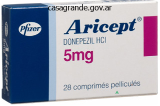
Aricept dosages: 10 mg, 5 mg
Aricept packs: 30 pills, 60 pills, 90 pills, 120 pills, 180 pills, 270 pills, 360 pills

Cheap 10 mg aricept amex
Neomycin (six per molecule), gentamicin (five), and tobramycin (five) produce the most renal damage; streptomycin (three) is the least. The function of molecular cost seems to be associated to binding of the cationic drug to receptors within the apical and subcellular membranes. At the apical membrane, the aminoglycoside may bind to anionic phospholipids, thereby promoting drug entry into the tubular cell. Within the cell, the aminoglycoside accumulates inside lysosomes, an impact that will also be dependent on cost. Inhibition of lysosomal capabilities (such as decreased synthesis of the proteolytic enzymes cathepsin B and cathepsin L) may then be answerable for the associated cellular damage. The likelihood of creating renal harm is dependent on the dose and the length of remedy. Prevention Careful monitoring of drug levels and minimizing the length of drug therapy are at present the first strategies used to reduce the incidence of aminoglycoside nephrotoxicity. Studies indicate that aminoglycoside nephrotoxicity can be minimized by giving the complete dose as quickly as a day (4 mg/kg in one study) quite than in three divided doses (1. Once-daily therapy ends in very high peak plasma and urinary concentrations; the latter exceeds the reabsorptive capacity of the proximal tubule so that many of the drug is excreted, not taken up by the tubular cells. The growth of distinction nephropathy in hospitalized sufferers is related to increased morbidity and mortality. Pathogenesis the mechanism of injury is believed to be a combination of direct vasoconstrictive results of the contrast agent with tubular toxicity mediated by technology of free radicals. Risk factors for creating contrast nephropathy embody current renal insufficiency, especially if a consequence of diabetic nephropathy, advanced coronary heart failure or different explanation for lowered renal perfusion (such as hypovolemia), high total dose of distinction agent and the osmolality of the agent used, and perhaps underlying multiple myeloma, especially if related to hypercalcemia. The renal damage sometimes begins on the day of the insult with hypotension or the administration of a radiocontrast agent; in comparison, the onset is delayed with aminoglycoside therapy. Recovery requires the regeneration of tubular cells, mediated in part by the activation of growth response genes and the release of development components. The recovery process can be accelerated in experimental animals; as examples, the administration of insulin-like progress factor I or epidermal growth issue can enhance tubular regeneration and the rate of improvement in renal function. The interval is shorter with a mild, self-limited damage and longer with a severe and chronic damage. For instance, patients with continued infection may have recurrent episodes of renal ischemia and prolonged renal injury, whereas patients with uncomplicated distinction nephropathy sometimes present peak creatinine concentrations at 3 to 5 days and then start to improve. It is due to this fact probably that the good majority of nephrons are poorly functioning or nonfunctioning at the time laboratory tests have been obtained. This course of is stimulated by a rise in the plasma potassium focus and by aldosterone (see Chapter 7). If, nonetheless, the plasma creatinine focus is secure for several days, then the steady state is present. This product reflects the amount of creatinine filtered and excreted, which, within the regular state, is the identical as the comparatively constant amount of creatinine produced. This will activate the tubuloglomerular suggestions system (see Chapter 1), which is ready to decrease the nephron filtration price till macula densa delivery has returned towards normal. Dissociation of tubular cell detachment and tubular cell dying in medical and experimental "acute tubular necrosis. She has developed a number of diabetic microvascular issues including retinopathy, peripheral neuropathy, and nephropathy. Albuminuria was first famous 15 years in the past, and since that time, protein excretion has progressively elevated to 5. Physical examination reveals a comparatively well-appearing, barely pale lady in no acute distress. Positive findings embrace a blood strain of 150/90 mmHg, decreased visible acuity bilaterally with proof of microaneurysms and exudates on funduscopic examination, 2+ (moderate) peripheral edema extending up to the midcalf, decreased vibration sense, and deep tendon reflexes. The role of parathyroid hormone and vitamin D in the normal regulation of calcium and phosphate stability.

Best order aricept
This oligoamnios was of placental origin as foetal origin was dominated out through focused scan for foetal malformations. The third was a spontaneous demise at 11 weeks after fetal coronary heart exercise was seen. This indicated that this time also the method of obstetric vasculopathy was in full swing. To wait, she wanted color Doppler for assuring the security of the foetus in the intrauterine surroundings. Any important alteration in the variability of the foetal heart rate and rhythm is ominous. Any a number of of those features indicate that the foetus is in bother and needs to be delivered. There are necessary inputs, which are obtained from the neonatologist, the primary being how early or preterm child will the neonatal unit be in a position to deal with. In this context, most good items are now moderately confident in handling the typical delivery weight of 1,000 g or extra. Many of those models are salvaging babies as small as seven-hundred g in weight and about 26 weeks in maturity. However, with better know-how, ventilator help and the availability of surfactants, nonetheless youthful and lighter babies will be salvaged. The second purpose to contain a neonatologist in this choice making is to help the unit get prepared for handling of such a child. It is, therefore, essential to contain the neonatologist in choice making relating to the timing of induction in pregnancies lower than 37 weeks in length increasingly more clear, it has turn out to be apparent that labour is induced not by the mother however by the foetal techniques. If the intrauterine environment is non-conducive to being pregnant continuation, labour will get induced rather more simply. Even if the foetus has had a demise, labour will get readily induced to get rid of the product of conception. Hence, a challenged and endangered foetal system in obstetric vasculopathy supports makes an attempt at labour induction readily. Also, the security profile turns into a matter of concern with this comparatively new pharmacological know-how. The Bishop score (also generally identified as pelvic score) is the most commonly used method to judge the readiness of the cervix for induction of labour. The Bishop rating gives factors to five measurements of the pelvic examination dilation, effacement of the cervix, station of the foetus, consistency of the cervix and position of the cervix. With the understanding of the physiology of labour becoming Methods of the induction of labour one hundred forty five Table 16. A rating of greater than 6 signifies a favourable cervix for induction of labour and will lead to a successful end result extra after than not. This is so because on extra events than not, labour inducing interventions have to be employed. For the success of any labour induction, cervical compliance and subsequent synchronous dilatation have to happen. Cervical ripening agents are anticipated to deliver about a sequence of changes within the ultrastructure and biochemical milieu in such a way that cervix turns into shortened, softened and dilated to let the passage of the foetus. This transforms the cervix from a sphincter organ, which preserves the being pregnant to a dilated conduit letting the foetus pass through it. Before the introduction of prostaglandins within the induction of labour, most pharmacological brokers like oxytocin used to act on the uterus. Their uterine predominance activated the exercise in the uterus secondary to which the cervix dilated. The uterine activity is going on in symphony with the cervical ripening and dilatation in rhythm. As a result, medication and methods that envisaged inducing labour by bringing about uterine exercise and letting the cervix respond secondarily to the uterus take a lengthy time for labour to get induced.
Order aricept 10 mg with amex
Immediate hypersensitivity Mechanism of Tissue Injury IgE antibodies-immediate release of vasoactive amines, lipid mediators, cytokines from mast cells- recruitment of inflammatory cells IgG, IgM antibodies; binding to target cell or matrix element: A. Autoimmune hemolytic anemia, erythroblastosis fetalis; membranous nephropathy, congenital membranous nephropathy, antibody-mediated graft rejection C. T cell mediated sarcoidosis, vasculitides (Takayasu aortitis, temporal arteritis), dermatomyositis, systemic sclerosis, inflammatory bowel disease, primary biliary cirrhosis, a quantity of sclerosis, autoimmune orchitis, granulomatous interstitial nephritis. This type of response is triggered when a divalent hapten or allergen cross-links immunoglobulin E (IgE) bound to specialised receptors for this immunoglobulin class on the floor of mast cells. Immune complexes kind in circulation elsewhere after which get trapped within the glomeruli, triggering lively irritation and eventually restore processes. Light microscopy in minimal change illness is basically regular or could reveal a gentle mesangial hypercellularity. Immunofluorescence microscopy (in which fluorescein-labeled antibodies directed against the different human immunoglobulins and other plasma proteins of curiosity are incubated with the kidney tissue) usually reveals solely "fine dusting" of IgG on or throughout the podocytes. The electron microscopy reveals marked degenerative modifications of the glomerular visceral epithelial cell, with diffuse retraction and effacement of foot processes, often referred to as "fusion" (arrows), microvillous degeneration of the cell surface (best seen in left upper segment), and presence of vacuoles (V) in the cytoplasm. The immunofluorescence microscopy often shows a "fine dusting" of immunoglobulin G over the podocytes but no deposits alongside the glomerular capillary walls (not shown). This sort of lesion represents the healed stage of a focal necrotizing and crescentic glomerulonephritis. Secondly, comparable adjustments can be induced in experimental animals by the administration of toxins corresponding to puromycin aminonucleoside or doxorubicin (Adriamycin), which affect predominantly the glomerular epithelial cells, or by antibodies specific for numerous cell floor elements of the visceral epithelium, including nephrin. However, relapses in minimal change illness often occur when remedy is discontinued. What stays unclear in human illness is the nature of the epithelial cell "toxin," also known as the "permeability factor. It is thought, for instance, that sufferers with end-stage kidney illness secondary to steroid-resistant nephrotic syndrome develop proteinuria after receiving a kidney allograft; these sufferers with familial disease have a defect within the gene that encodes for podocin, and during the posttransplant period, they develop antibodies directed in opposition to this epithelial cell surface protein related to the filtration slit diaphragm. The interaction between podocin and anti-podocin antibodies ends in abnormalities of the visceral epithelial cells that trigger proteinuria. A comparable state of affairs occurs in kids with congenital nephrotic syndrome after receiving a kidney transplant. These are examples of allo-reactive immune responses in patients naive to specific antigens present in the allograft. Similar diffuse epithelial cell damage with "fusion" of foot processes may additionally be induced in the course of the formation of immune complexes in the lamina rara externa (or subepithelial space), which is in close proximity to the epithelial cell membrane. This form of damage known as membranous glomerulopathy, nonetheless, is clearly antibody directed and complement mediated, as discussed within the following text. These illnesses are diffuse, and we can anticipate that every nephron is excreting abnormal ranges of albumin, which in flip results in sodium retention, edema, and finally the full nephrotic syndrome. Secondary Focal and Segmental Glomerulosclerosis It is important to appreciate that the light microscopic discovering of focal and segmental sclerosis is relatively nonspecific. During the therapeutic phase of any focal inflammatory, necrotizing, or ischemic glomerular damage as may occur in IgA nephropathy, lupus nephritis, or systemic polyangiitis, mentioned later in this chapter. These healed inflammatory lesions are greatest thought of focal and segmental glomerular scars. Genetic podocytopathies inflicting slowly progressive proteinuria or overt nephrotic syndrome. The lack of edema could be defined by the focal nature of the epithelial cell abnormality, which leads to elevated ranges of protein excretion and hence irregular sodium retention solely in few however not all nephrons. Immune Complex Formation the glomerulus is highly prone to the entrapment or native formation of immune complexes. The high plasma circulate rate (20% of the cardiac output), high intraglomerular strain, and excessive glomerular hydraulic conductivity which are required to promote filtration also increase the propensity for the deposition of antigens, antibodies, or antigen�antibody complexes. The subsequent development of an immune complex lattice could be detected as electron-dense aggregates by electron microscopy or as nice or coarse granules by immunohistochemistry (immunofluorescence microscopy or immune-enzymatic techniques) utilizing antibodies towards human immunoglobulin gentle and heavy chains.

Purchase generic aricept pills
Direct entry of rabies virus into the central nervous system without prior local replication. Reduced viral burden in paralytic in comparison with furious canine rabies is associated with outstanding inflammation at the brainstem level (In press). Intracellular unfold of rabies virus Is decreased within the paralytic form of canine rabies in comparability with the livid type. The distribution of challenge virus commonplace rabies virus versus skunk road rabies virus in the brains of experimentally infected rabid skunks. Street rabies virus causes dendritic harm and F-actin depolymerization in the hippocampus. Immunohistochemical examine of rabies virus within the central nervous system of domestic and wildlife species. Role of apoptosis in rabies viral encephalitis: A comparative research in mice, canine, and human mind with a evaluate of literature. Neuroanatomical mapping of rabies nucleocapsid viral antigen distribution and apoptosis in pathogenesis in street canine rabies-An immunohistochemical study. Rabies encephalitis following fox bite-Histological and immunohistochemical evaluation of lesions brought on by virus. Pathology of the peripheral nervous system in human rabies: A study of nine autopsy cases. Experimental rabies: Ultrastructural quantitative evaluation of the changes within the sciatic nerve. Apoptosis induction in mind in the course of the fastened pressure of rabies virus an infection correlates with onset and severity of illness. Laboratory methods in rabies: Rapid microscopic examination for negri bodies and preparation of specimens for biological take a look at. Rapid microscopic examination for Negri our bodies and preparation of specimens for organic take a look at. Neuronal dendritic morphology alterations within the cerebral cortex of rabies-infected mice: A Golgi examine (Spanish). Reactivation of Nedd-2, a developmentally down-regulated apoptotic gene, in apoptosis induced by a avenue pressure of rabies virus. Virus replication happens within the cell bodies and dendrites (Ugolini, 1995, 2010) from which newly fashioned viral particles are released (Bauer et al. After the an infection of muscle tissue cells and its entry into nerves, the virus has to address the innate immune response launched by the infected muscle and contaminated neurons which have the capability to counter the infection. Once infection is settled within the neurons, the infected neurons are shielded from the destruction by infiltrating T cells and by mechanisms limiting the inflammation of neuronal tissue. This virus invades the spinal wire and brain regions and causes fatal encephalitis (Camelo, Lafage, & Lafon, 2000; Park, Kondo, et al. Some of those receptors are on the surface of the cells, detecting the presence of danger signals current in the extracellular milieu. Recruitment of specific receptors depends upon the motifs that they bind to and the localization of the receptors. Viruses have developed sophisticated methods to escape the innate immune response (Randall & Goodbourn, 2008; Versteeg & Garcia-Sastre, 2010). The entry of the virus by the host is rapidly detected by host protection mechanisms in the periphery. This observation means that some viral particles may be readily eradicated at this early step of an infection. In the mind, each neurons and glial cells can mount antiviral, inflammatory, and chemokine responses. Nevertheless, an essential position is taken by microglia within the induction of neuroinflammation, a function which will replicate the density or the subcellular localization of the innate immune receptors (Bsibsi et al. These conditions ought to preserve not solely the integrity of the contaminated neuronal network, but additionally the lifetime of the host, permitting the virus to reach the brainstem and the salivary glands before the premature dying of the contaminated host.

Buy aricept 10mg on-line
Factors that trigger these nonspecific reactions are elusive, however one research recognized cross-reactive antibodies in acutely sick sufferers with other infectious illnesses (Rudd et al. For the aim of evaluating the oral vaccine baiting campaigns, willpower of particular person "protection" is much less essential than herd immunity ranges and the power to confidently compare results between laboratories and over time. The best rabies prevention applications embody rabies serology in varied steps, corresponding to registering and licensing new rabies vaccines, monitoring the immune status of people and animals, implementing animal vaccination campaigns, endeavor rabies surveillance and epidemiology, and, finally, sustaining rabies-free zones. It subsequently follows that selecting the best "fit-for-purpose" serological assay as nicely as guaranteeing that the take a look at is carried out correctly and that enough quality assurance procedures are in place is significant for the success of the program. Valid rabies serology outcomes affect worldwide trade agreements, the institution and upkeep of latest and current rabies-free zones, the everneeded development of new vaccines, and the analysis of research or surveillance outcomes printed in scientific journals. Performance traits are outlined by technique validation, which consists of an intensive plan of experiments to look at the accuracy, precision, specificity, sensitivity, 462 13. The experience of the technician depends on training and experience, and is proven by proficiency testing to reveal the flexibility to carry out the assay to outlined standards. The laboratory facility, including space, surroundings, and water quality, among different factors, must meet minimal criteria to guarantee that the testing is performed in a high quality managed state. Audits to establish standards and continual monitoring for the power to keep requirements is crucial for confidence within the check results produced. This is obtained through the use of only accredited reagents and supplies as well as only accredited tools as defined by the strategy validation. The pattern high quality as well as sample type are critical to the selection of the check technique in addition to the interpretation of the results. The laboratory must take steps to guarantee these components are addressed and all dangers are mitigated by way of a prime quality system implemented by the laboratory. The only method to know if a technique has the efficiency characteristics that "fit the aim" for which it will be used is to outline the check methodology via validation. A technique with acceptable accuracy and precision levels for the measurement of antibodies in a potency range of zero. The technique parameters important for a qualitative assay are sensitivity, specificity, and predictive value. In addition to sensitivity and specificity, a quantitative assay requires definition of accuracy (closeness to the true value), precision (repeatability of the measure), linearity, and reportable vary. Validations carried out are for specific purposes such as the evaluation of scientific trial samples for a human monoclonal antibody mixture for the postexposure therapy of rabies and vaccine potency analysis (Kostense et al. Robustness analysis describes the ability of the tactic to carry out to set criteria throughout normal variations in laboratory circumstances, including normal variations of apparatus efficiency, reagent tons, or between different personnel. Biologic variation (in sample and in biologic components of the assay) should be thought of separately from analytical variation. For instance, two take a look at outcomes from the identical pattern could vary solely on the basis of the receptivity of cells to virus an infection; cells used final week might have different virus infectivity characteristics than the cells utilized in subsequent testing. The variation from these types of factors is separate from other sources of variation. Interference can be brought on by cross-reacting antibodies, nonspecific binding, and the matrix impact (hemolysis, lipemia, or "dirty" samples, etc. Naturally occurring proteins in samples, similar to albumins, fibrinogen, and complement factors, can result in assay interference (Selby, 1999). When interference is suspected or needs to be dominated out, samples could additionally be evaluated by an alternate method by which the effect of interfering elements is minimized in order that specific exercise may be detected and measured. The trade-off is that a high cutoff level would improve the variety of false negatives. The probability of false optimistic and false negatives is said to the precision of the assay. Assays with a high variability significantly at the cut-off stage would exclude some true optimistic samples with potency values near the cut-off level and conversely identify some true negatives as positive. Upon repeat testing, these samples might generate both optimistic or adverse check results. Therefore, each time the sample matrix is altered, reevaluation of this parameter is required. Indeed, any change within the procedure or sample may require revalidation to decide the impact on the established efficiency characteristics. It is pure to evaluate the outcomes from different methods, and it is necessary to contemplate how the comparison is made. Although it is very common to consider settlement between strategies by a correlation coefficient, conclusions primarily based on this worth are improper.
Syndromes
- Meprobamate (Equanil)
- Yellow eyes
- Infection (a slight risk any time the skin is broken)
- You have had back pain before, but this episode is different and feels worse.
- Blurry vision
- Venous shunt surgery
- What drugs you are taking, even drugs or herbs you bought without a prescription

Order 5mg aricept free shipping
Underlying renal insufficiency is a crucial determinant of the degree of hyperkalemia in any form of hypoaldosteronism. Although relative lack of aldosterone decreases the effectivity of potassium secretion, sufferers with normal renal perform can compensate because a small rise in the plasma potassium concentration directly stimulates potassium secretion. Another speculation suggests that hypervolemia related to renal disease could be the major occasion. Volume enlargement increases levels of atrial natriuretic peptide that may suppress renin release. Aldosterone can directly stimulate distal hydrogen secretion, and decreasing aldosterone will promote the development of metabolic acidosis. However, hyperkalemia appears to be of greater importance as a end result of decreasing the plasma potassium concentration induces partial and even full normalization of the plasma bicarbonate concentration. Can you derive a mechanism by which hyperkalemia might cut back acid and ammonium excretion Consider the place the excess potassium will be distributed and how electroneutrality will be maintained. Symptoms the symptoms related to hyperkalemia are limited to muscle weakness (due to interference with neuromuscular transmission) and abnormal cardiac conduction. Disturbances in cardiac conduction induced by hyperkalemia can lead to cardiac arrest and dying. The earliest alteration is peaked and narrowed T waves due to more speedy repolarization; this change in T-wave configuration generally becomes apparent when the plasma potassium concentration exceeds from 6 to 7 mEq/L. Electrocardiogram in relation to the plasma potassium focus in hyperkalemia. The major exception happens in a disorder such as uncontrolled diabetes mellitus where the patient is actually potassium depleted and the elevation in plasma potassium concentration is as a end result of of a transcellular shift that can be reversed by remedy of the underlying disease. Because removing of potassium from the body will take time (through urinary excretion, gastrointestinal loss, or in extreme instances by dialysis; see following text), short-term treatment strategies involve temporary shifts of potassium from the extracellular to intracellular compartment. Dialysis Onset of Action Several minutes and then rapidly wanes Each of these modalities works within 30�60 minutes, lowers the plasma potassium focus by 0. Diuretics take a quantity of hours, however sufferers with advanced renal failure may show little response. General principles embrace avoidance of potassium supplements and discontinuation of medicine that may decrease aldosterone launch (see Table 7. The mostly used resin, sodium polystyrene sulfonate (Kayexalate), takes up potassium within the intestine and releases sodium. Indications for the opposite therapy modalities for hyperkalemia listed in Table 7. In this setting, insulin and glucose (10 models of insulin with at least forty g of glucose to stop hypoglycemia), a 2-adrenergic agonist (such as albuterol), and/or sodium bicarbonate may be given to transiently drive potassium into the cells till the excess potassium could be removed from the physique. As talked about at the beginning of this chapter, the preliminary depolarization of the resting membrane potential induced by hyperkalemia inactivates the sodium channels within the cell membrane leading to a decrease in membrane excitability. In patients with severe renal failure or on dialysis, acute dialysis may be indicated for potassium removal. Hypokalemia Hypokalemia is most frequently induced by elevated gastrointestinal or urinary losses, though elevated entry into the cell can also occur (Table 7. A low-potassium food plan alone generally has a comparatively minor effect on the plasma potassium focus as a outcome of urinary potassium excretion may be lowered to <15 to 25 mEq/day with potassium depletion. Increased entry into the cells-generally produces only a transient reduction in the plasma potassium focus A. Increased urinary losses-typically requires hyperaldosteronism and normal to enhanced distal flow A. Primary mineralocorticoid excess, most frequently as a outcome of aldosterone-producing adrenal adenoma D. Renal tubular acidosis In addition, potassium may be reabsorbed by the acid-secreting intercalated cells in the cortical collecting duct. The activity of this transporter is increased with potassium depletion (the sign for which may be a discount within the cell potassium concentration); the online effect is elevated potassium reabsorption and an appropriately decrease price of potassium excretion. The transcellular shifts and improve in urinary potassium excretion generally contain mechanisms much like however in the incorrect way from these described for hyperkalemia.
Discount aricept 5mg fast delivery
However, this patient has marked hypoalbuminemia, thereby reducing the focus of unmeasured anions. Thus, an anion gap of 15 mEq/L represents an elevation of 9 mEq/L, indicating the presence of a high anion gap metabolic acidosis. Physical examination reveals a blood pressure of 150/110 mmHg, and proximal muscle weak point is noted. The major causes of hyperkalemia, with particular emphasis on the importance of impaired urinary potassium excretion in sufferers with a persistent elevation within the plasma potassium focus. The physiologic rules that govern the selection of therapies for reversing hyperkalemia. The elements that can lower the plasma potassium concentration and the mechanisms by which urinary potassium losing can happen. Physiologic Effects of Potassium Total body potassium stores are approximately from three,000 to four,000 mEq. Roughly, 98% of the potassium is located within the cells; this distribution is in contrast to that of sodium, which is primarily limited to the extracellular fluid. First, it plays an necessary role in regulating a wide range of cell functions corresponding to protein and glycogen synthesis. Second, the ratio (rather than absolutely the values) of the potassium concentration within the cells ([K+]cell) to that within the extracellular fluid ([K+]ecf) is the most important determinant of the resting membrane potential (Em) throughout the cell membrane based on the following formulation: (Eq. Membrane excitability (or irritability) is equal to the distinction between the resting and threshold potentials; the latter is the potential during depolarization at which an motion potential is generated. Generation of the action potential is related to a marked elevation in sodium permeability, resulting in sodium entry into the cells and complete depolarization of the cell membrane. Changes within the plasma potassium concentration can have essential effects on membrane excitability. However, modifications in extracellular potassium have major effects on the state of activation of sodium channels. The web impact is increased sodium entry into cells, making Em much less unfavorable (closer to zero) and enhanced excitability that can lead to cardiac arrhythmias (see following discussion). Opposite adjustments are induced by an increase in the extracellular potassium concentration (hyperkalemia). The initial impact is to depolarize the membrane (make the potential less electronegative) and improve membrane excitability. This change, however, is transient because depolarization additionally tends to inactivate the sodium channels within the cell membrane. As with alterations in the plasma sodium focus (see Chapter 3), the likelihood of inducing symptoms with alterations in potassium balance is said both to the diploma and to the rapidity of change. As an example, the lack of potassium (as with severe diarrhea) will initially decrease the plasma potassium focus, has no impact on the cell potassium focus, and subsequently increases the ratio of mobile to extracellular potassium and make the resting potential extra electronegative. However, the autumn in the plasma potassium concentration creates a gradient that promotes potassium motion out of the cells; as this occurs, the concurrent discount in the cell potassium concentration leads to a smaller change within the ratio of mobile to extracellular potassium and therefore a lower probability of interfering with neuromuscular function and of inducing signs. In the steady state, the common potassium intake ranges from 40 to one hundred mEq/day (~1. These observations have essential implications for the regulation of potassium steadiness. The ingestion of 40 mEq of potassium (as with a number of giant glasses of orange juice) may, if the ingested potassium initially remained in the extracellular area, almost double the extracellular fluid potassium concentration (measured clinically as the plasma potassium concentration) and result in probably serious signs. Urinary potassium excretion, which is primarily determined by secretion within the principal cells within the cortical collecting tubule A. Plasma potassium focus Initial uptake of a few of the ingested potassium into the cells, thereby limiting the rise within the plasma potassium concentration. An understanding of the components that regulate these two steps is clinically essential because an abnormality in a single or both is present in many patients with an elevated plasma potassium focus (hyperkalemia) and in some sufferers with a low plasma potassium focus (hypokalemia). Potassium Uptake by Cells In normal subjects, three factors are of main significance in selling the transient movement of ingested potassium into the cells: a small elevation in plasma potassium concentration, insulin, and epinephrine (acting via the 2adrenergic receptors). The physiologic importance of these hormones has been demonstrated by the responses to a -adrenergic blocker. On the opposite hand, epinephrine launched throughout a stress response drives potassium into the cells and can transiently lower the plasma potassium concentration by as a lot as 1 mEq/L.

Buy generic aricept pills
Uric acid is excreted through kidneys, and so a high degree of uric acid meant impaired renal efficiency. Deli and Stefanovi showed that when parameters corresponding to uric acid (and urea) have been included to predict pre-eclampsia primarily based on the multivariate logistic regression model, they could correctly predict it in 79. The popularity of uric acid in pre-eclampsia was not confined to the realms of prediction. It can be an everyday entry on the record of "essential" investigations to be carried out in ladies being managed for pre-eclampsia in many massive and small hospitals. It is additional amusing that an undergraduate and postgraduate examinee is expected to mention uric acid estimation as an important investigation when asked to enumerate the listing of investigations in a case of pre-eclampsia. Sometimes it serves as a basic cell adhesion molecule by anchoring cells to collagen or proteoglycan substrates. Besides the prediction of pre-eclampsia, fibronectin has been studied extensively in predicting preterm labour. About 20 years ago, some research appeared that confirmed that plasma fibronectin levels could symbolize a particular marker for pre-eclampsia. Its high negative predictive worth strongly signifies that the event of pre-eclampsia is less probably in topics with high ranges inside the next four weeks after the blood sampling. It was discovered that single mid-trimester evaluation of fibronectin ranges in maternal plasma was not discovered to be helpful in predicting pre-eclampsia. Also, a technicality of assessment renders the testing of fibronectin impractical for mass screening. Subsequently, many instances algorithms are worked out to guide the clinician on how the clinician has to move along for analysis and administration of pre-eclampsia. Nearly all of the high- and moderate-risk factors have been studied individually or in numerous combinations for prediction of pre-eclampsia. The authors have instructed that estimation of such a priori danger for pre-eclampsia is an important first step in the use of Bayes theorem to combine maternal (epidemiological) Colour doppler in pre-eclampsia prediction: the sport changer 71 factors with biomarkers for the continuing improvement of a more effective technique of screening for pre-eclampsia. X-rays were the one precept imaging modality for medical science until as late as the early 1980s. What began as a non-invasive imaging approach to study the blood provide and its complexities has become a potent weapon for efficient prediction, prognostication and determination making in different stages of various obstetric vasculopathies, together with pre-eclampsia. For the prediction of pre-eclampsia, uterine artery colour Doppler is nicely researched. Uterine arteries could be readily accessed to colour Doppler as a end result of it crosses the iliac vessels earlier than getting into the uterine musculature and just after giving the cervical (descending) branch. A substantial threat of issues associated with impedance to maternal adjustments on the level of uterine circulation has been demonstrated. It was attention-grabbing to examine the diastolic notch and the behaviour of indices at around 12 weeks after which once more at or around mid-trimester. Basically, pulsatility index, resistance index and systolic-todiastolic ratio assist us in identifying the quantum of diastolic circulate within the uterine vascular system. This may be better understood if correlated with the pathophysiology occurring at or round this time. The strategy of the second wave of trophoblastic invasion is expected to get competently accomplished at or round mid-trimester. With this moulding, the maternal vascular bed turns into a low-resistance, high-flow pool of blood. It becomes practically shielded off from the opposite adjustments which are taking place within the maternal methods. It also gets shielded off from the pressor substances that might be in circulation in the maternal system at a while or the other. The sum total impact of all these changes is a rise in blood flow to the pregnant uterus. That apart, this increase in blood flow is continuous and guarded by blunting the effects of sympathetic and parasympathetic systems on spiral arterioles. In the earlier days of software of color Doppler in the obstetric world, there was no settlement on the index or a mix of indices to be used for predicting adverse situations of preeclampsia. They scanned each uterine arteries at round 20�24 weeks in high-risk subjects for pre-eclampsia. They discovered that the measurement of serum Activin-A and Inhibin-A levels could add important prognostic information for predicting pre-eclampsia in pregnant girls exhibiting specific Doppler alterations late in the second trimester. In a evaluate article printed in 2015, a total of six biomarker algorithms, for the prediction of pre-eclampsia had been studied.

Buy aricept toronto
This limitation to gentle microscopy is essential because most glomerular ailments contain almost the entire glomeruli if the latter are examined by electron or immunofluorescence microscopy. Global-Involving the complete glomerular tuft; could be seen with either focal or diffuse illness. Glomerulonephritis-Any condition related to inflammation in the glomerular tuft. Nephrotic versus Nephritic Syndromes the 2 cases introduced firstly of this chapter illustrate the attribute scientific manifestations of the two major glomerular syndromes: nephrotic and nephritic. The last abnormality primarily displays elevated hepatic lipoprotein synthesis and decreased catabolism induced in an unknown method by the fall in plasma oncotic strain (which is primarily determined by the plasma albumin concentration). Clinical Manifestations, Structural Patterns of Injury, and Mechanism of Glomerular Diseases Clinical Manifestation Nephrotic syndrome Structural Pattern of Injury 1. Direct cytokine impact; experimentally, direct effect of poisons or following antibody binding to the visceral epithelial cell parts 3. Diffuse and international glomerulosclerosis Rapidly progressive glomerulonephritis Hematuria/proteinuria 8. Podocyte dysfunction and structural abnormalities also can result from defects in the genes that encode for proteins of key structural components of the podocyte (the filtration slit diaphragm, the cytoskeleton, cell adhesion equipment, etc. The lack of glomerular inflammation and subsequently of extreme acute tissue damage explains two different features of the scientific presentation of the nephrotic syndrome: the urine sediment is comparatively inactive, containing few cells or mobile casts, and the plasma creatinine focus is often regular or solely mildly elevated at presentation. The widespread structural finding in all nephrotic circumstances is distinguished and in depth damage of the glomerular visceral epithelial cells manifested by diffuse simplification or effacement of foot processes, also commonly referred to as "fusion" of foot processes. Because the primary target of the harm is the glomerular visceral epithelial cell, the podocyte, these situations can be considered as main podocytopathies. Nephritic Syndrome Although heavy proteinuria also can occur in nephritic states, the characteristic scientific discovering is an energetic urine sediment containing purple cells (sometimes with gross hematuria), white cells, and cellular and granular casts, as in Case 2. The prominent urinary abnormalities in this setting replicate the inflow into the glomerular tuft of circulating inflammatory cells together with neutrophils, macrophages, monocytes, and generally lymphocytes. The primary site and goal of the injury in these situations are components throughout the more proximal layer of the capillary wall, namely, the endothelium and the lamina rara interna. The kind and severity of the glomerular inflammation determines the level of the kidney dysfunction and related scientific manifestations (see Table 9. Patients with extreme glomerular injury involving most or the entire glomeruli with energetic irritation present with a variable and normally sudden elevation in the plasma creatinine concentration. The sudden discount within the rate of glomerular filtration in diseases with diffuse involvement of glomeruli can also induce sodium retention within the distal tubule, leading to extracellular fluid quantity growth, edema, and hypertension. These patients normally current with a constellation of signs and symptoms that represent the acute nephritic syndrome. [newline]Other inflammatory diseases result principally in a focal necrotizing and/or crescentic sample of glomerular injury. This syndrome known as quickly progressive glomerulonephritis to differentiate it from the extra sudden acute nephritic syndrome; the signs within the latter are normally noticed in a single day. Another group of diseases presents solely with focal and often segmental irritation of the glomeruli; such processes sometimes present with asymptomatic hematuria, delicate and even no proteinuria, and a normal plasma creatinine focus and systemic blood stress. A fifth glomerular syndrome, continual kidney failure, is characterized by slowly progressive loss of function over many months or years, typically associated with rising proteinuria and variable hematuria; superior glomerular, tubulointerstitial, vascular, and systemic illnesses with kidney harm can end result in this constellation of indicators and signs, often additionally referred to as end-stage kidney disease. There are four major mechanisms by which the glomerular inflammatory process and the nephritic state could be induced; these are totally different from the mechanisms responsible for the nephrotic presentation: Immune advanced formation and complement activation in the subendothelial space or in the mesangium as happens in poststreptococcal glomerulonephritis, other infection-associated glomerulonephritides, immunoglobulin A (IgA) nephropathy, and in some patients with lupus nephritis; circulating monoclonal immunoglobulins associated with Bcell lymphoproliferative problems, plasma cell dyscrasias, or multiple myeloma can also induce this kind of tissue damage. Activation of complement through the alternative pathway; the dysregulation of complement activation is because of genetic defects of the components of this pathway or acquired functional interference at various steps of this process by autoantibodies or paraproteins. The widespread structural finding in all nephritic circumstances is injury of the endothelium that results in lively irritation because of antibody deposition or binding and/or complement activation in shut proximity to this cell or immune complex formation and deposition within the subendothelial and mesangial areas. The attainable function of cell-mediated immunity stays uncertain in most glomerular and vascular illnesses; nevertheless, this mechanism is clearly responsible for some interstitial nephritides and a few forms of allograft rejection. The the rest of this chapter discusses these mechanisms of glomerular and vascular illness. It is helpful, nevertheless, to begin with a short evaluation of glomerular construction and performance, which helps to explain how proteinuria would possibly occur and the websites at which immune deposits are likely to type. These sites are an necessary determinant of the type of illness that may ensue; as noted previously, subepithelial immune deposits lead to epithelial cell injury and a nephrotic presentation, whereas mesangial or subendothelial immune deposits sometimes result in irritation of the glomerulus and to a nephritic presentation. The outermost layer of the capillary wall is made up of the visceral epithelial cells or podocytes. Adjacent foot processes are derived from totally different epithelial cells and are connected to each other by modified desmosomes often known as the filtration slit diaphragms. The glomerular capillary community is connected and arranged around a central zone, the mesangium.

Order aricept 10 mg on line
Serial sampling of suspect patients is perfect for definitive diagnosis and have to be thought of earlier than rendering a unfavorable prognosis utilizing this pattern type alone. If the affected person remains alive but unwell for more than 2�3 weeks or recovers rapidly, the differential prognosis is less likely to include rabies. Interestingly, however, it has been noted that sufferers with paralytic rabies may give false-negative outcomes by such checks, maybe because of the timing of sample collection and/or the very restricted titers of virus current in these sample sorts in patients exhibiting this form of the illness (Hemachudha & Wacharapluesadee, 2004; Wacharapluesadee & Hemachudha, 2002). Such issues reinforce the necessity for pan-lyssavirus diagnostic tests, particularly for those patients who present within the developed world and for whom world journey is frequent. Although efficiency of antemortem diagnosis in home animals has not often been thought of, animal rights considerations seeking to keep away from pointless euthanasia of animals for rabies testing might exert stress to develop sturdy animal antemortem testing regimens in the future. Without advanced supportive medical care, a rabid animal will succumb to the illness quickly. By the time diagnostic outcomes could be available on a battery of samples similar to those described for people, the animal would most probably be moribund or lifeless. Moreover, negative findings early in the clinical course of a rabid animal could mislead public well being administration of doubtless uncovered individuals. Thus, in most techniques that rely on passive submissions for diagnostic testing, the true incidence of the illness is unknown and finally demonstrating its elimination by way of control programs is troublesome. Moreover, as canine rabies continues to flow into in many international locations the potential for its reintroduction, by way of translocation of diseased "rescue canine," into developed countries during which the disease has been eliminated, has been described in several reports (Hercules et al. Of course, the impression of rabies on human health stays the principal driver toward elimination of this illness as a end result of the large prices that it can confer onto public well being techniques as properly as the emotional distress that the disease imparts. A single rabies case in the wrong place, similar to a county fair or pet shop, can lead to the triage of large numbers of individuals for potential publicity to this animal and the necessity for postexposure prophylaxis (Noah et al. Despite the clear public well being threat, fiscal constraints usually jeopardize implementation of the infrastructure critical to rabies prevention, a problem especially severe for governments of creating international locations. Accordingly, continued development of dependable, accurate, cost-effective, and timely main diagnostic tools is needed in the fight to fight this disease. Clarifying indeterminate outcomes on the rabies direct fluorescent antibody test using real-time reverse transcriptase polymerase chain reaction. Pathobiological investigation of naturally infected canine rabies cases from Sri Lanka. Distribution of rabies antigen in contaminated mind material: Determining the reliability of different areas of the mind for the rabies fluorescent antibody check. Molecular strategies to distinguish between classical rabies and the rabies-related European bat lyssaviruses. Rapid detection of rabies virus by reverse transcription loop-mediated isothermal amplification. Phylogenetic relationships amongst rhabdoviruses inferred using the L polymerase gene. Antemortem analysis of human rabies in a veterinarian contaminated when handling a herbivore in Minas Gerais, ~ Brazil. Susceptibility of u sheep to European bat lyssavirus type-1 and-2 infection: A medical pathogenesis examine. The long incubation interval in rabies: Delayed progression of an infection in muscle on the web site of publicity. An inter-laboratory proficiency testing train for rabies diagnosis in Latin America and the Caribbean. Reverse transcription recombinase polymerase amplification assay for fast detection of canine related rabies virus in Africa. Dual mixed realtime reverse transcription polymerase chain reaction assay for the diagnosis of lyssavirus infection. Clinical and epidemiological features of human rabies circumstances in the Philippines: A evaluation from 1987 to 2006. Evaluation of six commercially available fast immunochromatographic checks for the analysis of rabies in brain material. Revue scientifique et approach (International Office of Epizootics), 37(2), 421�437. Molecular doubleu examine technique for the identification and characterization of European lyssaviruses. Emerging technologies for the detection of rabies virus: Challenges and hopes in the 21st century.
References
- Greenstein A, Plymate SR, Katz PG: Visually stimulated erection in castrated men, J Urol 153:650n652, 1995.
- Knobler H, Savion N, Shenkman B, et al: Shear-induced platelet adhesion and aggregation on subendothelium are increased in diabetic patients. Thromb Res 1998;90: 181-190.
- Wilt TJ, Jones KM, Barry MJ, et al. Follow-up of prostatectomy versus observation for early prostate cancer. N Engl J Med 2017;377(2):132-142.
- Ferreri AJ, Montalban C. Primary diffuse large B-cell lymphoma of the stomach. Crit Rev Oncol/Hematol 2007;63:65.
- Oral H, et al. Catheter ablation for paroxysmal atrial fibrillation: segmental pulmonary vein ostial ablation versus left atrial ablation. Circulation 2003;108:2355-2360.
- Kaouk, J.H., Haber, G.P., Goel, R.K. et al. Single-port laparoscopic surgery in urology: initial experience. Urology 2008;71:3-6.
- Lyons JA, Kupelian PA, Mohan DS, et al. Importance of high radiation doses (72 Gy or greater) in the treatment of stage T1-T3 adenocarcinoma of the prostate. Urology 2000;55(1):85-90.
- Braunwald E, Brown WV, eds. Atlas of heart disease: vascular disease, Vol. 10.


