David Childs, MD
- Assistant Professor of Radiology
- Abdominal Imaging Section
- Wake Forest University School of Medicine
- Winston-Salem, North Carolina
Careprost dosages: 3 ml
Careprost packs: 1 bottles, 2 bottles, 3 bottles, 4 bottles, 5 bottles, 6 bottles, 7 bottles, 8 bottles, 9 bottles, 10 bottles
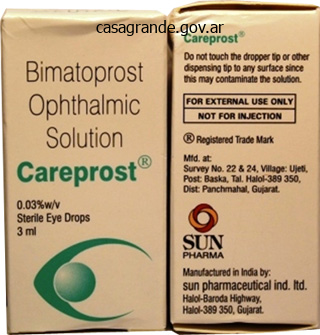
Buy on line careprost
Because the clever surface permits the harvest of aligned cells as a single-cell sheet, cell sheet engineering overcomes these limitations to facilitate the design of 3D cell orientation inside a densely packed cell environment. To achieve this, several sorts of micropatterned thermoresponsive substrates have been developed [91,92]. Using these substrates, cell orientation is first regulated 2D after which the aligned cells are stacked as cell sheets layer by layer. Microgrooved polydimethylsiloxane substrates are extensively used to control cell alignment, and the same method can be utilized to produce aligned cells on the thermoresponsive surface [93]. As a end result, the floor features to regulate cell orientation and release the cell sheet. The width of the patterns is a key issue within the regulation of cell orientation, and the earlier examine demonstrated that a 50 � 50-mm striped sample was acceptable for producing a cell sheet composed of aligned cells. Because of the distinction in the cell-to-surface affinity on the surfaces, cells are aligned parallel to the stripe patterns simply by cell seeding. This suggests that the cell orientation influences the bodily properties of cell sheets. In addition, the cell alignment induces a change within the organic properties of cell sheets. Adapted with permission from Takahashi H, Nakayama M, Shimizu T, Yamato M, Okano T. Anisotropic cell sheets for setting up three-dimensional tissue with well-organized cell orientation. Therefore, this could potentially enhance vascularization in multilayered cell sheets. Arrangement of Three-Dimensional Orientation Using Cell Sheet Layering Techniques Because cell sheets can be manipulated using a gelatin gel-coated plunger [53], cell sheets composed of aligned cells can be layered while sustaining the designed orientation. In some native tissues, tissue anisotropy is organized 3D and the orientation of their buildings is essential for specific mechanical and biological capabilities. For instance, native myocardial tissue consists of multiple layers of aligned cardiomyocytes which are oriented in various instructions throughout the entire tissue [88,89,96]. This well-organized 3D oriented construction is necessary for producing the anisotropic electrical propagation discovered in the native myocardium. However, it remains difficult to arrange totally different cell orientations vertically using typical biomaterial scaffolds. This strategy is easy and versatile for creating a selection of tissue architectures. As described earlier, the cell-dense tissue structure is important for multiple cell sheets to communicate with each other within the layered cell sheet assemble. Therefore, this technique of arranging cell orientation inside engineered tissues is expected to be helpful for producing biomimetic myocardial tissues. Skeletal Muscle Tissue Engineering Because native skeletal muscle tissue is made from extremely oriented myofibers, the orientation of muscle cells needs to be regulated to produce biomimetic muscle tissue constructs [97e99]. Adapted with permission from Takahashi H, Shimizu T, Nakayama M, Yamato M, Okano T. The use of anisotropic cell sheets to control orientation during the self-organization of 3D muscle tissue. After reaching confluence, the aligned myoblasts could be harvested as a single steady cell sheet by lowering the culture temperature to 20 C. In addition, regulating the aligned orientation stimulates the formation of longer myotubes, in contrast with randomly oriented myoblasts. This aligned construction will be important for producing biomimetic muscle tissue [100]. Moreover, as a result of the aligned myotubes may be layered with cell sheet know-how, this mixture helps the manufacturing of 3D aligned muscle tissues, which is required for scaling up skeletal muscle tissues. Consequently, all myoblasts grew to become aligned throughout the layered cell sheet construct. Furthermore, this self-organizing conduct permits us merely to produce 3D myotube constructs with a single orientation.
Proven careprost 3ml
Collagen orientation in periosteum and perichondrium is aligned with preferential directions of tissue growth. A microgroove patterned multiwell cell tradition plate for highthroughput studies of cell alignment. Cell sheet-based tissue engineering for organizing anisotropic tissue constructs produced using microfabricated thermoresponsive substrates. A thermoresponsive, microtextured substrate for cell sheet engineering with outlined structural organization. Micropatterned thermoresponsive polymer brush surfaces for fabricating cell sheets with well-controlled orientational structures. Multifunctional cell-culture platform for aligned cell sheet monitoring, transfer printing, and therapy. Scalable alignment of three-dimensional cellular constructs in a microfluidic chip. The affect of electrospun aligned poly(epsilon-caprolactone)/collagen nanofiber meshes on the formation of self-aligned skeletal muscle myotubes. Tissue regeneration could be achieved by stimulating the repair mechanisms of the body, particularly by the supply of stem cells that can perform biological features and materials that may function as scaffolds to promote cell proliferation. In addition, many functional nanomaterials have been used to deliver drugs, proteins, and genes for tissue regeneration [4]. Since Richard Feynman first proposed the idea of growing expertise at the atomic scale in 1959, nanotechnology has been quickly developed and actively investigated in lots of biomedical research fields. We may also introduce nanotechnology-based stem cell remedy, significantly for controlling stem cell fate and performance. In neural tissue, the axon preliminary section is responsible for generating motion potentials in neurons and is organized at the nanoscale. This structure is organized periodically in a longitudinal direction and is layered radially. The sarcomeres consist of protein filaments composed of actin and are organized in a course parallel to thick filaments composed of aggregates of myosin. In tendon tissue, a tendon fiber is composed of a hierarchical construction of tendon fibers, fascicle, and nanoscale collagen fibrils (50e500 nm), that are additional composed of collagen molecules (w1. To fabricate biomimetic scaffolds that can be used to enhance tissue regeneration, nanomaterials such as nanoparticles, nanofibers, and nanofilms have been developed to mimic the composition and structure of tissues at the nanoscale [4,5]. Physical Properties the physical properties of natural systems, similar to their size, shape, and surface topography, are instantly associated to their function. Nanogrooved buildings can be fashioned on substrates to promote the alignment of cardiomyocytes for cardiac tissue engineering. This tissue polarity may be recapitulated on nanofibers by surface modification to enhance cell adhesion. In addition, the surface texture of cell substrates, significantly on the nanoscale, can influence varied cellular processes similar to differentiation, motility, spreading, and apoptosis [13]. For instance, epithelial cells grow in an elongated type on surfaces patterned with nanoscale grooves and ridges, whereas these cells have a rounded morphology on easy surfaces [11]. These observations have promoted the event of nanomaterials with novel bodily properties for purposes in regenerative drugs. The small size of nanoparticles results in favorable properties compared with microparticle-based delivery strategies, such as improved circulation and the flexibility to cross tissue limitations [15]. The size of the nanoparticle is correlated with the efficiency of mobile uptake and is set by the avidity of ligandereceptor interactions between the nanoparticle and the cell surface [16]. However, nanoparticles of sizes 50 nm or larger can bind a number of receptors but can restrict the extra binding of nanoparticles. These properties can differ in accordance with the cell type and tradition conditions, which might cause variations in the number of receptors and internalization mechanisms [16]. These nanomaterials can self-assemble into a larger structural unit based on covalent or noncovalent interaction. For instance, peptides with an amphiphilic structure can self-assemble into tubular constructions.
Buy careprost from india
The geometry of the underlying substrate was also found to have an result on fibroblast attachment, during which the greatest adhesion occurred at ridges however diminished on nanosized pits and columns [116]. Differences in proliferation and adhesion can lead to differential endocytotic activities of cells on topographies. Human fibroblasts attempted to endocytose nanocolumns 100 nm in diameter fabricated using colloidal lithography [117]. The enhanced mobile endocytosis exhibited by the cells on microand nanopillars promoted the internalization of fluorescently labeled dextran, in addition to the nonviral gene transfection of green-fluorescent protein encoding plasmid with lipofectamine. By optimization and careful design, varied topographies can be applied to regulate drug and gene supply and cell proliferation. Corneal endothelial cells typically have low in vitro proliferation rates and are nonproliferative in vivo. However, studies show that acceptable surface topographical cues can induce higher proliferation rates in corneal endothelial cells [119] as well as in main human corneal endothelial cells [120]. Pathological adjustments to the microstructure of basement membranes can occur underneath pathological situations. This model was subsequently used to examine the monolayer formation and migration of major corneal endothelial cells on guttata-like microtopography [121]. This examine is an example of how in vitro disease models can be utilized to mimic basement membrane microstructural adjustments and consequently to research the mechanisms underlying cell habits in pathological settings. Whereas many studies have demonstrated the significant affect of substrate topography on mobile responses, researchers have additionally been actively investigating the mechanisms underlying the topographical regulation of cell habits. It has long been demonstrated that genetic expression can be influenced by surface topography. A lowered expression of integrin subunits a2, aV, b2, b3, and b4 was noticed on nanopatterned substrates in contrast with unpatterned substrates. Actomyosin contractility could be one other intracellular mechanism concerned in topography-induced differentiation. Another study recognized zyxin as one of the molecular players concerned in the topography-mediated modulation of mobile response [126]. Interestingly, a higher turnover rate of zyxin was observed when cells had been cultured on more compliant substrates [127]. Therefore, phenomenologically, the preliminary molecular response of a cell to nanotopography seems to mirror that of a compliant substrate. Another essential part of the mechanistic understanding of topography-induced differentiation is the effect on the stem cell differentiation efficiency and velocity, and the essential time interval for making use of topography to improve stem cell differentiation. The group also compared the importance of a priming interval by which the stem cells had been cultured on the topography in maintenance media, with the actual contact of topography through the differentiation interval. The study demonstrated that topography contact is critical for topography-induced differentiation, and the impact of topography priming and topography contact is additive in enhancing differentiation. In addition to stem celledirected differentiation, a research confirmed that topography at the microscale can promote the direct conversion of fibroblasts into neurons [129]. The purity and conversion effectivity of the induced neurons (iN) were increased on micrograting substrates, whereas neurite branching was elevated on microposts and decreased on microgratings. Although that examine sheds gentle on the impression of topography on the cytoplasmic stage, another study linked extracellular physical cues to changes in the nucleus [130]. Electrically Conductive Substrate Bioelectricity is the electrical currents and electrical potentials generated by or occurring within residing cells, tissues, and organisms. Similarly, cardiac tissue consists of aligned cells and cardiomyocytes, which conduct the electrical present that gives indicators for cardiac contraction and blood move. The heart consists of Purkinje fibers, that are electrically conductive and essential for the conduction system of the guts. Because electrical current is an indispensable a half of cell and tissue operate, electrically conductive substrates and scaffolds for tissue engineering applications can be designed to facilitate higher mobile cross-talk and tissue regeneration. This limitation may be addressed to an extent by mixing conductive polymers corresponding to polyaniline and polypyrrole with the scaffold materials answer earlier than processing.
Discount careprost online master card
Stem cell and biomaterials research in dental tissue engineering and regeneration. Role of nanostructured biopolymers and bioceramics in enamel, dentin and periodontal tissue regeneration. Self-assembling peptide amphiphile nanofibers as a scaffold for dental stem cells. Nanoparticle-based bioactive agent launch systems for bone and cartilage tissue engineering. New insights into and novel applications of launch know-how for periodontal reconstructive therapies. Antibacterial activity of chitosan nanofiber meshes with liposomes immobilized releasing gentamicin. Dual launch of a hydrophilic and a hydrophobic osteogenic issue from a single liposome. Immobilization of bioactive factor-loaded liposomes on the floor of electrospun nanofibers focusing on tissue engineering. Instructive nanofibrous scaffold comprising runt-related transcription factor 2 gene supply for bone tissue engineering. Decellularized scaffolds: ideas, methodologies, and purposes in cardiac tissue engineering and whole-organ regeneration. Characterization of natural, decellularized and reseeded porcine tooth bud matrices. Odontogenic induction of dental stem cells by extracellular matrix-inspired three-dimensional scaffold. Tooth root regeneration utilizing dental follicle cell sheets together with a dentin matrix based scaffold. Periodontal therapeutic by periodontal ligament cell sheets in a tooth replantation mannequin. Regeneration of dental pulp/dentine advanced with a three-dimensional and scaffoldfree stem-cell sheet-derived pellet. Generation of a bioengineered tooth by using a three-dimensional cell manipulation methodology (organ germ method). Tooth germ-like assemble transplantation for whole-tooth regeneration: an in vivo examine within the miniature pig. Three dimensional dental epithelial-mesenchymal constructs of predetermined size and form for tooth regeneration. Tissue engineering of complicated tooth structures on biodegradable polymer scaffolds. The sequential seeding of epithelial and mesenchymal cells for tissue-engineered tooth regeneration. Fibrin glue combined with platelet-rich fibrin as a scaffold seeded with dental bud cells for tooth regeneration. The interaction of dental pulp stem cells and endothelial cells in an injectable peptide hydrogel on angiogenesis and pulp regeneration in vivo. Effects of hyaluronic acid sponge as a scaffold on odontoblastic cell line and amputated dental pulp. In vivo technology of dental pulp-like tissue through the use of dental pulp stem cells, dentin matrix protein 1 transplantation in mice. Scaffoldless tissue-engineered dental pulp cell constructs for endodontic remedy. Regeneration of periodontal tissues: mixtures of barrier membranes and grafting materials - organic basis and preclinical proof: a scientific review. Periodontal ligament stem cell-mediated treatment for periodontitis in miniature swine. Periodontal regeneration with multi-layered periodontal ligamentderived cell sheets in a canine mannequin. Mesenchymal stem cell traits of dental pulp and periodontal ligament stem cells after in vivo transplantation. A platelet-derived growth issue releasing chitosan/coral composite scaffold for periodontal tissue engineering. Reconstructing mandibular defects using autologous tissueengineered tooth and bone constructs.
Order careprost 3ml line
Maternal T cells limit engraftment after in utero hematopoietic cell transplantation in mice. High-level allogeneic chimerism achieved by prenatal tolerance induction and postnatal nonmyeloablative bone marrow transplantation. In utero hematopoietic stem cell transplantation: ontogenic opportunities and biologic barriers. Induction of tolerance in nondefective mice after in utero transplantation of main histocompatibility complex-mismatched fetal hematopoietic stem cells. In utero bone marrow transplantation induces donor-specific tolerance by a combination of clonal deletion and clonal anergy. Hemopoietic chimerism in rodents transplanted in utero with fetal human hemopoietic cells. In utero hematopoietic stem cell transplantation: progress toward scientific application. The maternal immune response inhibits the success of in utero hematopoietic cell transplantation. Regulation of the earliest immune response to in utero hematopoietic cellular transplantation. Prenatal transplantation of cytokine-stimulated marrow improves early chimerism in a resistant strain mixture however ends in poor long-term engraftment. Maternal administration of busulfan before in utero transplantation of human hematopoietic stem cells enhances engraftments in sheep. In utero depletion of fetal hematopoietic stem cells improves engraftment after neonatal transplantation in mice. Gestational age of recipient determines pattern and degree of transgene expression following in utero retroviral gene transfer. In utero gene therapy: transfer and long-term expression of the bacterial neo(r) gene in sheep after direct injection of retroviral vectors into preimmune fetuses. Rescue of enzyme deficiency in embryonic diaphragm in a mouse model of metabolic myopathy: Pompe illness. Lentivirus-mediated gene transfer to the developing bronchiolar airway epithelium in the fetal lamb. Sustained phenotypic correction of canine hemophilia A utilizing an adeno-associated viral vector. Clinical and molecular characterization of a re-established line of sheep exhibiting hemophilia A. Cost-effectiveness in establishing hemophilia Carrier detection and prenatal prognosis companies in a growing country with restricted well being sources. Induction of secure prenatal tolerance to beta-galactosidase by in utero gene transfer into preimmune sheep fetuses. Permanent phenotypic correction of hemophilia B in immunocompetent mice by prenatal gene therapy. Early fetal gene supply makes use of both central and peripheral mechanisms of tolerance induction. In utero transduction of hematopoietic cells is enhanced at early gestational ages. Fetal gene switch using lentiviral vectors and the potential for germ cell transduction in rhesus monkeys (Macaca mulatta). Factors figuring out the risk of inadvertent retroviral transduction of male germ cells after in utero gene transfer in sheep. Male germ-line cells are at risk following direct-injection retroviral-mediated gene transfer in utero. Transduction of long-term-engrafting human hematopoietic stem cells by retroviral vectors. Long-term luciferase expression monitored by bioluminescence imaging after adeno-associated virus-mediated fetal gene supply in rhesus monkeys (Macaca mulatta). Lentiviral vector gene switch into fetal rhesus monkeys (Macaca mulatta): lung-targeting approaches. Rhesus monkey model for fetal gene switch: research with retroviralbased vector techniques. Reexpression following readministration of an adenoviral vector in grownup mice after preliminary in utero adenoviral administration.
Buy careprost 3 ml online
Worldwide, every year approximately 15 million individuals experience a stroke (World Heart Federation). For all of those conditions, acute and persistent populations pose different treatment challenges. In contrast, the transport of bigger molecules must be actively facilitated; nutrients. The inflammatory response is initiated by peripherally derived immune cells such as macrophages and activated glial cells such as microglia that migrate into the harm website. Reactive Astrocytes and the Glial Scar Under normal situations, glial cells in the mind and spinal twine have a giant number of necessary roles, together with upkeep and regulation of the extracellular microenvironment [10]. Astrocytes secrete numerous trophic elements that help neurons, regulate extracellular ranges of ions and neurotransmitters, and provide an antioxidant defense for neurons [11]. In the retina, Muller cell gliosis involves the upregulation of important intermediate filaments and the expression of neuroprotective cytokines and factors that encourage photoreceptor survival after harm [20]. Further work is needed to establish tips on how to steadiness each roles of reactive astrocytes successfully to promote wound healing while facilitating axon regeneration. Endogenous Stem Cells Spontaneous host tissue regeneration is dependent upon endogenous cells with the capacity to proliferate and differentiate. Neural stem cells have also been found in the spinal wire, particularly within the ependymal layer of the subependymal zone of the central canal [24,25], as well as inside the parenchyma after injury [25a]. Retinal stem cells have been identified in the pigmented ciliary epithelium of the attention; they proliferate in vitro and are differentiated into retinal-specific cell sorts in mice [27] and people [28]. Differences in wound response, anatomy, and cell kind necessitate therapies tailored to the tissue goal and pathological state, whether or not with biomolecule or cellbased therapeutics. Local injections could be performed into the epidural or intrathecal spaces of the spinal wire, into the ventricles in the mind, or instantly into the tissue. Neuroprotection can be outlined as a long-lasting upkeep of practical ability, in addition to increased axonal sparing and reduced lesion volume. Neuroprotective molecules act on quite so much of mobile targets, similar to antiapoptotic pathways to reduce death of certain neuronal populations or oligodendrocytes, and antiinflammatory pathways to inhibit microglial activation [32]. Growth elements beforehand explored for his or her regenerative potential have also been shown to be neuroprotective. Neuroregeneration consists of the expansion of existing axons, sprouting of new axons from neural cell soma, remyelination, and plasticity amongst surviving axons, all resulting in tissue restore and practical recovery. Blocking this interplay with anti-NogoA or anti-NgR allows neurite outgrowth to resume [44]. Another method to neuroregeneration is stimulation of endogenous stem and progenitor cells within the stem cell area of interest. Stem cells ameliorate poststroke impairments within the mind by modulating inflammation, inducing angiogenesis, and secreting neuroprotective components into host tissue [52e54]. A main problem with transplanting allogeneic or xenogeneic cells is a lack of long-term transplant survival, likely the results of the hostile microenvironment of the injury [58e60]. An different technique involves the directed differentiation of stem cells, which have the flexibility for unlimited development in culture. The inner mass cells proliferate and differentiate to become the cells that make up the three germ layers of creating humans with the potential to become any cell within the physique (pluripotent). This propagation potential could be recapitulated in culture and differentiation of those cells may be directed in a lineage-specific method to yield terminally differentiated cells [61]. Although there are issues that their proliferative capacity might lead to tumor and mass formation [62], none have been reported clinically. Predifferentiation of those cells is a way to diminish the implied danger of instantly transplanting pluripotent cells [63]. This trial was suspended twice in the first yr: as soon as because of questions concerning the purity of the cell inhabitants and then owing to microscopic cysts in the regenerating damage web site in animal fashions. The comparatively poor survival of transplanted cells means that a mixture of cells and drug/biomolecule supply would improve survival and integration of the transplanted cell inhabitants. In terms of morbidity and mortality, it is doubtless one of the most typical neurological problems.
Cheap careprost 3 ml line
Injectable self-gelling composites for bone tissue engineering primarily based on gellan gum hydrogel enriched with completely different bioglasses. Enzymatic mineralization of gellan gum hydrogel for bone tissue-engineering functions and its enhancement by polydopamine. Composites of gellan gum hydrogel enzymatically mineralized with calcium-zinc phosphate for bone regeneration with antibacterial exercise. Enrichment of enzymatically mineralized gellan gum hydrogels with phlorotannin-rich Ecklonia cava extract Seanol((R)) to endow antibacterial properties and promote mineralization. Endothelial differentiation of human stem cells seeded onto electrospun polyhydroxybutyrate/polyhydroxybutyrate-co-hydroxyvalerate fiber mesh. Tuning the properties of polyhydroxybutyrate films utilizing acetic acid through solvent casting. Preparation of poly(3-hydroxybutyrate)/nano-hydroxyapatite composite scaffolds for bone tissue engineering. Porous 3d implants of degradable poly-3hydroxybutyrate used to improve regeneration of rat cranial defect. Antibacterial nanostructured polyhydroxybutyrate membranes for guided bone regeneration. Biocomposite scaffolds based mostly on electrospun poly(3hydroxybutyrate) nanofibers and electrosprayed hydroxyapatite nanoparticles for bone tissue engineering purposes. Development of silk-based scaffolds for tissue engineering of bone from human adipose-derived stem cells. Electrospun silk fibroin/poly(lactide-co-epsilon-caprolactone) nanofibrous scaffolds for bone regeneration. Hierarchically porous nagelschmidtite bioceramic-silk scaffolds for bone tissue engineering. Bioactive macro/micro porous silk fibroin/nano-sized calcium phosphate scaffolds with potential for bone-tissue-engineering functions. De novo bone formation on macro/microporous silk and silk/nano-sized calcium phosphate scaffolds. Bilayered silk/silk-nanoCaP scaffolds for osteochondral tissue engineering: in vitro and in vivo assessment of biological performance. Physicochemical properties, modifications and functions of starches from totally different botanical sources. Design and characterization of a biodegradable double-layer scaffold aimed at periodontal tissue-engineering purposes. Porous starch/cellulose nanofibers composite prepared by salt leaching approach for tissue engineering. A novel bidirectional continuous perfusion bioreactor for the tradition of largesized bone tissue-engineered constructs. Presence of starch enhances in vitro biodegradation and biocompatibility of a gentamicin supply formulation. Nano-hydroxyapatite/chitosan-starch nanocomposite as a novel bone assemble: synthesis and in vitro studies. Undifferentiated human adipose-derived stromal/stem cells loaded onto wet-spun starch-polycaprolactone scaffolds enhance bone regeneration: nude mice calvarial defect in vivo examine. The impact of differentiation stage of amniotic fluid stem cells on bone regeneration. Evaluation of a starch-based double layer scaffold for bone regeneration in a rat mannequin. Micro-structured calcium phosphate ceramic for donor website repair after harvesting chin bone for grafting alveolar clefts in youngsters. Cutaneous and labyrinthine tolerance of bioactive glass S53P4 in mastoid and epitympanic obliteration surgery: prospective clinical research. Quantitative evaluation of the regenerative and mineralogenic performances of the zebrafish caudal fin. Comparative research of hydroxyapatite from eggshells and synthetic hydroxyapatite for bone regeneration. Natural-based nanocomposites for bone tissue engineering and regenerative medicine: a evaluation. A porous hydroxyapatite scaffold for bone tissue engineering: physico-mechanical and organic evaluations.
Order careprost canada
In flip, these can be particularly separated from other nucleated cells by their adherence properties to plastic culture flasks. The synergistic profit of these immunomodulatory effects is a reduction in the danger for host rejection. Scaffolds are sometimes subdivided into three lessons based mostly on their material composition: polymers, metals, and bioceramics. Synthetic scaffolds have the benefit of typically being able to be fabricated with a wider vary of degradation rates and mechanical properties compared with their pure counterparts. This capability is based on their polymeric composition, the copolymer ratio, and the interactions of their polymeric side chains. A novel enviornment within scaffold design has been the adoption of additive manufacturing applied sciences (3D printing) in scaffold fabrication. The time period "3D printing" describes a bunch of additive manufacturing applied sciences that are collectively referred to as solid free-form fabrication. These embody laser stereolithography, fiber electrospinning, fused deposition modeling, fiber extrusion from the soften, and injection molding [21]. The advantages of 3D printing are conferred by its immense flexibility in fabricating scaffolds of varying structural complexity. This course of allows for quite so much of control over the assemble architecture and suppleness in scaling up fabrication, and it has the added benefit of technically precise reproducibility, which is usually lacking in subtractive fabrication strategies. The interaction between signaling molecules and growth components is advanced and multivariable. Factors critical to cell signaling embrace the spatiotemporal release of progress elements and their bioactivity. The supply of biochemical signaling cues is usually categorized as unbound, sure inside the implant with a designed managed supply, coated on the implant surface, or coded within the cells by way of gene delivery mechanisms [23,24]. Unbound delivery systems are marked by the rapid efflux of the expansion elements adopted by rapid clearance from the microenvironment. Bound supply techniques permit the benefit of managed or specific variable launch of the biochemical signal over time. Hydrogels are the most generally investigated polymer for cell encapsulation and the in situ delivery of biochemical cues [22]. Marshall Urist discovered that demineralized bone matrix induced ectopic bone formation in subcutaneous and intramuscular pockets in rodents [27]. There are a quantity of drug supply systems under investigation that permit for measured controlled delivery. Among other supply methods, these permit for the temporal and spatial release of progress factors and cell alerts in a controlled fashion. In focusing on specific cascades, investigators are extra capable of understanding specific variable responses in a closed setting with the objective of predicting future responses in preclinical animal models and in subsequent translational first-in-human purposes. In vitro studies afford the chance to simulate complex interactions amongst cells, scaffolds, and growth elements in comparatively controlled environments. Bone-forming cells (osteoblasts) are sometimes procured from three primary origins: � pluripotent stem cells that differentiate into osteoblasts [31], � primary osteoblasts and osteocytes from completely different species [32,33], and � immortalized cell strains [34,35]. To maintain translational validity, bone in vitro fashions should incorporate the traditional triad of cells, scaffolds, and development factors in a fashion that intently mimics both the biochemical and biomechanical bone formation characteristics that are seen in vivo. Historically, the study of the interaction between bone physiological and pathological processes has been carried out on 2D plastic plates. In vitro studies provide nice perception into the translational validity of a mannequin, but the biomechanical environment in vivo is much more complex and the stresses exerted on the bone dictate its resorption and deposition charges. In Vivo Preclinical Models the benefit of in vivo fashions rests in the capacity to assess biomaterials in more complicated tissue environments which have variable loading conditions. It has been well-illustrated that bone composition, density, and mechanical properties of various animal models. Chief among these is that the animal model demonstrates physiological and pathophysiological processes that mimic those in people. The animal administration and care points should have been thought-about and are optimal for the chosen study. Selection Considerations Based on Animal Species Animal species which are into consideration for in vivo preclinical research embody mice, rats, rabbits, sheep, goats, pigs, dogs, cats, and nonhuman primates. Mice and rats are available and cheap and have minimal housing necessities.
References
- Adams HP Jr, del Zoppo G, Alberts MJ, et al: Guidelines for the early management of adults with ischemic stroke: a guideline from the American Heart Association/American Stroke Association Stroke Council, Clinical Cardiology Council, Cardiovascular Radiology and Intervention Council, and the Atherosclerotic Peripheral Vascular Disease and Quality of Care Outcomes in Research Interdisciplinary Working Groups: the American Academy of Neurology affirms the value of this guideline as an educational tool for neurologists, Circulation 115:e478-e534, 2007.
- Effects of calcium antagonists on the risks of coronary heart disease, cancer and bleeding. Ad Hoc Subcommittee of the Liaison Committee of the World Health Organisation and the International Society of Hypertension. J Hypertens. 1997; 15(2):105-115.
- Foroud T, Koller DL, Lai D, et al. Genome-wide association study of intracranial aneurysms confirms role of Anril and SOX17 in disease risk. Stroke 2012;43(11):2846-52.
- Pow-Sang J, Ojeda J, Ramirez G, et al: Carcinoma of the penis: analysis of 192 consecutive cases at the Instituto Nacional de Enfermedades Neoplasicas, Int Adv Surg Oncol 2:201n221, 1979.


