
Harry Snyder
- Lecturer, Health Policy and Management

https://publichealth.berkeley.edu/people/harry-snyder/
Losartan dosages: 50 mg, 25 mg
Losartan packs: 28 pills, 56 pills, 112 pills, 224 pills, 30 pills, 60 pills, 90 pills, 120 pills, 180 pills, 270 pills, 360 pills
Losartan 25mg sale
Currently the pathogenic mechanism is believed to contain a disordered regenerative course of following damage to the alveolar epithelium and the apoptosis of type I pneumocytes induced by an exterior agent. Cytokines activate the discharge of myofibroblasts, which Distant metastasis M0 M1 No distant metastasis Distant metastasis, together with separate tumor nodule (s) in a contralateral lobe 152 Downloaded by: Tulane University. The smooth margins and popcorn calcifications within the tumor matrix are typical of hamartoma. In the setting of normal tissue regeneration, the myofibroblasts would eventually turn into apoptotic after serving their operate. The prognosis is very poor, with a imply survival of two to four years, so that early consideration of lung transplantation is beneficial. The adjustments show an inhomogeneous distribution with a marked gradient from the central to peripheral lung and from base to apex. This is accompanied by subpleural parenchymal destruction making a honeycomb sample of cystic structures which might be sometimes three to 10 mm in diameter however might attain 2. The intrapulmonary fibrotic foci lead to traction bronchiectasis, and total lung volume is lowered. The mobile form is characterized by the histological predominance of an interstitial mobile inflammatory response and has a considerably better prognosis. Smoking-Related Idiopathic Interstitial Pneumonias Bronchiolitis-Associated Interstitial Lung Disease Brief definition. The pigmentation is attributable to granular inclusions from compounds current in cigarette smoke. Even the superior stage shows no honeycomb pattern and minimal traction bronchiectasis. The alveolar walls are thickened due to infiltration by inflammatory cells (eosinophilic granulocytes and lymphocytes). Desquamated alveolar cells (type I pneumocytes) are also found in the alveolar area. Smoking cessation is the best remedy, and that measure alone will result in medical and radiological improvement. Acute and Subacute Idiopathic Interstitial Pneumonias Cryptogenic Organizing Pneumonia Brief definition. It is characterized histologically by buds of granulation tissue with myofibroblasts and fibrocytes together with intercellular matrix within the alveolar airspace. The prognosis is very good, and most cases will resolve utterly with corticosteroid remedy. A particular feature is the "reverse halo sign," which may be seen in 20% of patients. It is produced by a ringlike, barely ragged opacity that surrounds normal tissue or parenchyma of ground-glass opacity. In the late stage, pulmonary fibrosis results in traction bronchiectasis and the formation of a honeycomb pattern. Severe dyspnea develops over a period of a quantity of weeks in a beforehand wholesome affected person or following a viral infection. The dyspnea progresses rapidly to respiratory failure requiring mechanical ventilation. The predominance of infiltrates within the perilymphatic tissue leads to thickening of the interlobar septa and bronchovascular bundle. In approximately 80% of patients, thin-walled perivascular cysts from a quantity of millimeters to 30 mm in diameter are found in the central lung. Rare Idiopathic Interstitial Pneumonias Lymphoid Interstitial Pneumonia Brief definition. There could also be a secondary accumulation of macrophages and protein-rich fluid within the alveoli. The hallmark of the illness is the development of fibrosis alongside the pleural floor including the interlobar fissures. If the acute stage is survived, this late stage is variable and ranges from good restoration of pulmonary structure to honeycombing and fibrosis with excessive cellularity and a small proportion of collagen fibers. Differentiation is especially required from prior tuberculosis with apical pleural caps.
Cheap losartan 50mg with visa
Lower lobe atelectasis can also be associated with elevation of the ipsilateral hemidiaphragm. The medical presentation is decided by the disease underlying the atelectasis. In patients with absorption atelectasis brought on by a tumor, the main scientific findings are B signs (undesired weight loss, fever, night sweats) and four four. The entities overlap in ailments similar to pulmonary sequestration and pulmonary artery aplasia. The medical spectrum is decided by the severity of disease and might vary from onset of signs in maturity to respiratory misery in the new child. Bilateral pulmonary agenesis is incompatible with postnatal life, however the majority of cases are unilateral. Associated cardiac anomalies are common, and tracheoesophageal fistula may be present. The affected hemithorax is smaller than the contralateral aspect, and tons of children will develop scoliosis. The chest radiograph exhibits opacity of the affected hemithorax with overinflation of the contralateral lung. Aplasia Absent proximal segment Interruption, congenital absence Hypoplasia Stenosis 4 Pulmonary arteries Sling formation Variants in vessel origins (Truncus arteriosus) Arteriovenous fistulas Arteriovenous connections Arteriovenous malformations Pulmonary veins Anomalous pulmonary venous return Anomalous pulmonary venous drainage Scimitar syndrome Intralobar sequestration Downloaded by: Tulane University. In unilateral cases, possible pulmonary hypertension will decide the prognosis. Cystic adenomatoid malformation of the lung results from abnormal budding of the terminal bronchi early in the first trimester of being pregnant. This results in an overgrowth of bronchial tissue, which, being derived from the terminal bronchioles, is devoid of cartilage. The dysplastic bronchial constructions might have a connection to the tracheobronchial tree, by which case the cysts can drain and should appear as ring shadows on chest radiographs. It may be traced in continuity from that region to the pulmonary apex, with extension into the mediastinum. Bronchogenic cysts could be very difficult to distinguish from cystic adenomatoid lung malformation and, when massive, might produce the same set of signs. Congenital lobar emphysema refers to the overinflation of one pulmonary lobe or section. Possible causes are a check-valve mechanism as a end result of circumscribed bronchomalacia (a wall cartilage defect might result in expiratory collapse with an obstructive check-valve mechanism), bronchial stenosis, or presence of a mass, or may be idiopathic. The left upper lobe is affected in 50% of cases, while the proper higher and middle lobes each account for about 20% of circumstances. Pronounced overinflation might cause mediastinal shift and the atelectasis of adjacent, unaffected pulmonary segments. The malformed tissue is normally confined to one lobe and will produce a mass impact causing compression of the esophagus and trachea and a mediastinal shift. Malignant transformation with the formation of varied kinds of sarcoma or bronchoalveolar carcinoma is known to happen, and therefore resection is the treatment of selection even for smaller lesions. Due to the pathogenic mechanism described above, the disease becomes clinically apparent solely with the onset of respiration after start. Approximately 50% of kids manifest dyspnea and cyanosis during the first month of life. In milder types of the anomaly, the diagnosis could additionally be instructed by recurrent episodes of pneumonia in the course of the first months of life. The identification of vascular markings in congenital lobar emphysema permits differentiation from pneumothorax and congenital cysts. Congenital lobar emphysema may result from a check-valve mechanism with inspiratory overinflation of the affected lobe or section. The left upper lobe is affected in onehalf of instances, and the proper upper and middle lobes are every affected in 20%. The cause is normally a circumscribed cartilage defect in the bronchus to the affected lobe, destabilizing the bronchial lumen and resulting in circumscribed bronchomalacia. Affected youngsters show early symptom onset with dyspnea and cyanosis, usually enabling the situation to be recognized in the course of the first months of life. Only a small percentage of patients have recurrent episodes of pneumonia as their dominant symptom. The diploma of pulmonary hypoplasia determines the severity of postnatal respiratory distress.
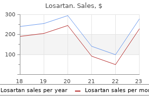
Buy losartan 50mg lowest price
In the best hand there are streaky areas of sclerosis of the little finger, affecting the metacarpal and phalanges. This patient has the monomelic kind with involvement of only the proper upper limb. Streaky and patchy sclerosis is present within the scapula and extends along all of the tubular bones. Includes: Osteopathia striata (isolated); osteopathia striata with pigmentary dermopathy; Voorhoeve illness. Occasionally cardiac anomalies, talipes, postaxial polydactyly, reasonable studying difficulties, seizures, hypothyroidism. Rarely extreme problems could happen associated to moyamoya or to cervical backbone instability. Sclerosis of the bottom of the skull; poorly aerated sinuses; sclerosis of mastoid air cells. Regular, fantastic, longitudinal metaphyseal strains of elevated density often extending to the diaphyses; fan-like striations of the iliac bones. Females might current with macrocephaly, cleft palate, and delicate learning disabilities and have regular life expectancy. Narrowing of the neural foramina on the base of the cranium might lead to cranial nerve deficits together with deafness, recurrent facial nerve palsy, and ophthalmoplegia. Surviving affected males are likely mosaics and have increased bone density with much less apparent striations. Surgery when indicated for cleft palate, maxillary hypoplasia, and cervical backbone stabilization. Melorheostosis: the sclerosis is thicker, uneven, and extra irregular than in osteopathia striata. Sponastrime dysplasia is differentiated by midface hypoplasia with a depressed nasal bridge and the biconcave vertebral our bodies, in addition to brief stature of affected individuals. Enchondromatosis: Oval-shaped lytic lesions and tumefactions are found on this situation however not in osteopathia striata. Goltz-Gorlin syndrome: Longitudinal bands of decreased radiodensity separated by osseous septa are seen on this X-linked dominant. This disorder factors to frequent pathogenic mechanisms causing osteopathia striata, melorheostosis, and osteopoikilosis. Cortina H, Vallcanera A, Vidal J (1981) Familial osteopathia striata with cranial condensation. In addition to sclerosis of the skull progressive sclerosis and thickening of the frontal and occipital bones is seen. The short tubular bones show some undermodeling and there are fine striations within the distal radii. Prenatally, sonography could show nuchal edema and short limbs at 12 weeks gestation. Advanced skeletal maturation, notably of the carpal and tarsal bones and laryngeal cartilages. Short, thick ribs giving a slender thorax and short thick upwardly curved clavicles. This accelerates the differentiation of chondrocytes and subsequent bone formation, explaining the elevated bone mass and untimely ossification. Kniest dysplasia differs by the much less severe shortening of the tubular bones, regular bone density, regular ribs, and irregular vertebral our bodies. Painful, agency, gentle tissue swelling involving significantly the cheeks, jaws, scapular region, arms, and legs; often with pseudoparalysis of a limb. In later levels of protracted courses, bowing and/or elevated length of the concerned long bones; delayed motor development. Short-limb quick stature and intrauterine or perinatal death in extreme instances; polyhydramnios could additionally be associated.
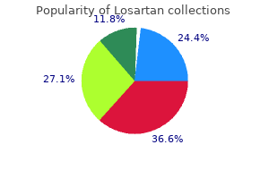
Best losartan 50 mg
The disease occurs predominantly in 5- to 10-year-old boys, and a limp is normally the rst medical sign. Bones that comprise red bone marrow are the extra common metastatic sites (spine, cranium, ribs, pelvis, and femora). It is the most typical type of arthritis and may be thought-about a normal part of the getting older course of. As the condition worsens, joints become less mobile, and new growths of cartilage and bone are seen as osteophytes (bony outgrowths). Pelvic ri g fractures: Because of the closed ring structure of the pelvis, a severe blow or trauma to one side of the pelvis might result in a fracture site away from the location of major trauma, thus requiring clear radiographic visualization of the complete pelvis. Proxim al fem ur (hip) fractures: these fractures are most typical in older grownup or geriatric patients with osteoporosis or avascular necrosis. Both osteoporosis (loss of bone mass from metabolic or other factors) and avascular (loss of blood circulation) necrosis (cell death) frequently result in weakening or collapse of weightbearing joints such as the hip joint; fractures happen with only minimal trauma. Slipped capital fem oral epiphysis (S F): this condition normally occurs in 10- to 16-year-olds throughout fast growth, when even minor trauma can precipitate its growth. The epiphysis appears shorter and the epiphyseal plate wider, with smaller margins. Routine to embrace both joints: Common departmental routines include both joints on all preliminary femur exams. If the hip is included, the leg ought to be rotated 15� to 20� internally to place the femoral neck in pro le. Po sitio n: n o rotatio is evidenced; femoral and tibial � condyles ought to seem symmetric in dimension and form with the outline of patella barely toward medial side of femur. Exp o su re: Optimal publicity with right use of anode heel � impact or use of compensating lter will lead to close to uniform density (brightness) of complete femur. Po sitio n, e the ra l: Anterior and posterior margins of Tru La � medial and lateral femoral condyles should be superimposed and aligned with open patellofemoral joint area. Pa rt sitio n Po � Flex affected knee about 45� and align femur to midline of table. Positio n, e the ra l: Superimposition of the greater and Tru La � lesser trochanters by the femur exists, with solely a small a part of the trochanters seen on medial side. Femur must be centered to collimation eld with hip joint a minimum of 1 inch (2. Exp o su re: Optimal exposure with right use of anode heel � impact or use of compensating lter will end in near-uniform density (brightness) of whole femur. Correct centering evidenced by demonstration of entire pelvis and superior femora with out foreshortening in collimated eld. Exp o su re: Optimal publicity visualizes L5 and sacrum space � and margins of the femoral heads and acetabula, as seen by way of overlying pelvic constructions, with out overexposing the ischium and pubic bones. Shield gonads for each males and females without obscuring essential anatomy (see Note 1). Po sitio n: n o rotatio is evidenced by symmetric � look of the pelvic bones, particularly the ala of the ilium, two obturator foramina, and ischial spines, if seen. Exp o su re: Optimal exposure visualizes the margins of the � femoral head and the acetabulum by way of overlying pelvic constructions, with out overexposing the proximal femora. Po sitio n: n o rotatio: Obturator foramina and bilateral � ischia are equal in measurement and shape. Exp o su re: Body and superior rami of pubis are well � demonstrated without overexposure of ischial rami. Gonadal shielding is feasible for males if care is taken not to obscure important pelvic anatomy. Po sitio n: n o rotatio: Ischial spines are absolutely demonstrated � and equal in size and shape. Proper centering and angulation are evidenced by demonstration of the superimposed anterior and posterior parts of the pelvic ring. Exp o su re: Optimal exposure demonstrates the � superimposed anterior and posterior parts of the pelvic ring. Pa rt sitio n Po � Place patient in 45� posterior indirect, with both pelvis and thorax 45� from tabletop.
Diseases
- Myopathy Moebius Robin syndrome
- Setleis syndrome
- MILS syndrome
- Mental retardation coloboma slimness
- Hypercalcemia, familial benign type 3
- Anophthalia
Purchase losartan without prescription
The ilia wings are crescent-shaped, the decrease portions of the ilia are hypoplastic, and the acetabular roofs are broad and horizontal. There is a shallow, inverted-V-shaped configuration of the expansion plates of the distal femora. The proximal ends of the femora are hyperplastic and the capital femoral epiphyses are small. The metaphyses are mildly flared with irregular margins and the epiphyses are flattened. Metacarpals and phalanges are quick with flared metaphyses and irregular epiphyses. On both sides of the physeal strains, the bone structure is grossly irregular with "flocky" ossification, clear and wide metaphyses, and dysplastic epiphyses. Acute bowing of the distal portion of the femoral shaft results in a striking genu valgum. The sample of bone modifications is similar to that in the severe phenotype with flared long bone ends, waver-thin vertebral bodies, and brief ilia with halberd-shaped crests. However, hyperplasia of the rib ends, vertebral pedicles, and metaphyses is much less conspicuous than within the extreme phenotype. The vertebral our bodies are flat, and the basilar portions of the ilia are underdeveloped. In both patients, marked platyspondyly is current but no kyphoscoliosis has developed. The vertebral our bodies are flat and anteriorly pointed with irregular higher and lower plates. The vertebral our bodies are flat with a bony buildup within the posterior two-thirds of the lumbar bodies. At all ages, the lower ilia are hypoplastic with broad, horizontal acetabular roofs. The capital femoral epiphyses of the youngsters and the femoral heads of the adult are small and the trochanteric area is plump. Bone structure is irregular in A and C however only mildly paying homage to the popcorn aspect seen within the parastremmatic phenotype. In this patient the hand bones had been severely affected and indistinguishable from those in typical metatropic dysplasia. However, a diagnosis could also be delayed as a end result of short stature is absent or gentle in infancy and early childhood. Broad, flared ilia with short larger sciatic notches and horizontal acetabula; the iliac change is mild to almost normal in milder phenotypes. Normal to mildly flared metaphyses of the lengthy bones; metaphyseal flaring may be conspicuous in youthful youngsters with severer phenotypes. Kyphoscoliosis and C1/C2 instability might result in myelopathy which will require neurosurgical and orthopedic intervention. Morquio disease differs by the anterior tonguing of the vertebral our bodies, hypoplasia of the decrease ilia, fine corneal opacities, and keratansulfaturia. The vertebral bodies are elongated with a mild overfaced pedicles look on the upper lumbar backbone. The vertebral bodies are flattened, rectangular, and elongated with an overfaced pedicles look. Waddling gait in early childhood, restricted joint mobility; occasionally genua valga. Progressive kyphoscoliosis; typically manifesting in early childhood, frequent in adolescence and maturity. Generalized platyspondyly with some anterior wedging, kyphosis, and scoliosis of varying degree. Broad, short basilar parts of the iliac bones, with broad horizontal acetabular bones and horizontal acetabular roofs (more marked in children than in adults). Irregular metaphyseal ossification in tubular bones, normally most marked within the proximal femora the place coxa vara are frequent. Cesarean part could additionally be required in affected females due to narrowing of the pelvic outlet.
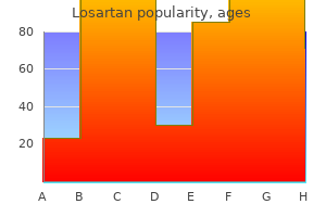
Purchase generic losartan line
L � Elbow should be exed 90� in true lateral place, as evidenced by direct superimposition o epicondyles. These, along with accompanying lecture slides o radiographs, present a basis or classroom or positioning laboratory discussion. Critique these radiographs or errors in a number of o the f ve categories, as described on this textbook and as outlined on the best. Cla vicle S ca pula Hume rus H the hum erus is the biggest and longest bone o the higher limb. The anatomy o the distal humerus and o the elbow joint was described in Chapter 4. The barely constricted space instantly under and lateral to the pinnacle is the anat m ic neck, which appears as a line o demarcation between the rounded head and the adjoining higher and lesser tubercles. The process directly below the anatomic neck on the anterior sur ace is the lesser tubercle (tu-ber-kl). The bigger lateral course of is the larger tubercle, to which the pectoralis major and supraspinatus muscle tissue attach. The deep groove between these two tubercles is the intertubercular (in-ter-tu-ber-ku-lar) sulcus (bicipital groove). The tapered space under the pinnacle and tubercles is the surgical neck, and distal to the surgical neck is the lengthy b dy (sha t) o the humerus. The delt id tuber sity is the roughened raised triangular elevation alongside the anterolateral sur ace o the physique (sha t) to which the deltoid muscle is hooked up. Some anatomic elements are more di f cult to visualize on radiographs than on drawings. However, an excellent understanding o the situation and relationship between varied components helps in this identif cation. The unction o the clavicle and scapula is to connect each higher limb to the trunk or axial skeleton. Anteriorly, the shoulder girdle connects to the trunk at the higher sternum; nonetheless, posteriorly, the connection to the trunk is incomplete because the scapula is linked to the trunk by muscles only. Each shoulder girdle and every higher limb connect on the shoulder joint between the scapula and the humerus. The higher margin o the scapula is on the degree o the sec nd p steri r rib, and the lower margin is at the degree o the seventh p steri r rib (T7). Cla vicle the clavicle (collarbone) is an extended bone with a double curvature that has three main components: two ends and a long central portion. The lateral or acr m ial (ah-kro-me-al) extrem ity (end) o the clavicle articulates with the acromion o the scapula. The medial or sternal extrem ity (end) articulates with the manubrium, which is the upper part o the sternum. This joint also is well palpated, and the mixture o the sternoclavicular joints on either facet o the manubrium helps to orm an necessary positioning landmark referred to as the jugular (jug-u-lar) n tch. The b dy (sha t) o the clavicle is the elongated portion between the 2 extremities. The acromial finish o the clavicle is attened and has a downward curvature at its attachment with the acromion. The sternal finish is extra triangular in shape, broader, and is directed downward to articulate with the sternum. The male clavicle tends to be thicker and more curved, normally being most curved in closely muscled males. The lateral angle, generally referred to as the head of the scapula, is the thickest part and ends laterally in a shallow melancholy called the glenoid cavity (ossa). The humeral head articulates with the glenoid cavity o the scapula to orm the scapul hum eral (skap-u-lo-hu-mer-al) j int, also called the glenohumeral joint, or shoulder joint. The superi r and inferi r angles re er to the upper and decrease ends o the medial or vertebral border. The anterior sur ace o the scapula is termed the c stal (kostal) floor as a end result of o its proximity to the ribs (costa, literally which means "rib"). The center area o the costal sur ace presents a large concavity or despair, generally identified as the subscapular f ssa.
Buy genuine losartan on-line
Exp o su re: Optimal density (brightness) and contrast with � n m ti n reveal clear, sharp bony trabecular markings. Exp o su re: Optimal density (brightness) and contrast with � n m ti n reveal clear, sharp bony trabecular markings and pertinent so t tissue anatomy. I the position is per ormed with the patient within the recumbent position, place supports under elevated shoulder and hip to keep this place. Exp o su re: Optimal density (brightness) and distinction with � n m ti n visualize so t tissue margins and clear, sharp bony trabecular markings. Exp o su re: Optimal density (brightness) and contrast with � n m ti n visualize sharp borders and sharp bony trabecular markings and reveal the entire intertubercular sulcus seen by way of so t tissue with out extreme density. Exp o su re: Optimal density (brightness) and contrast with � n m ti n visualize sharp bony trabecular markings and pertinent so t tissue anatomy. This technique will blur the surrounding pulmonary buildings while preserving the proximal humerus in a relatively stationary position. Exp o su re: Optimal density (brightness) and contrast � show whole define o the humeral head and the proximal hal o the humerus. Exp o su re: Optimal density (brightness) and distinction with � no movement visualize sharp bony borders and the outline o the physique o the scapula by way of the proximal humerus. Po sitio n: Acromion and coracoid processes ought to appear � as practically symmetric upper limbs o the "Y � the humeral. Exp o su re: Optimal density (brightness) and contrast � demonstrate the Y appearance o the higher lateral scapula superimposed by the humeral head with outline o the physique o the scapula visible via the humerus. Because o di erences among sufferers, the quantity o physique obliquity may vary rom 45� to 60�. Po sitio n: the coracoid process is projected over half o the � humeral head, which seems elongated. Exp o su re: Optimal density (brightness) and distinction with � n m ti n reveal clear, sharp bony trabecular markings and so t tissue detail or potential calcif cations. Exp o su re: � Midclavicle, sternal, and acromial extremities show clear, sharp bony trabecular markings and so t tissue element. Less weight (5 to 8 lb per limb) could also be used or smaller or asthenic sufferers, and more weight could also be used or larger or hypersthenic patients. Holding onto weights might end in alse-negative radiographs as a end result of they have an inclination to pull on the weights, resulting in contraction somewhat than relaxation o the shoulder muscle tissue. W G: this technique should be used only by skilled and qualif ed personnel to forestall further harm. Po sitio n: A ected arm seen to be abducted ninety levels � and hand supinated, as evidenced by the lateral border o the scapula ree o superimposition. Exp o su re: Optimal density (brightness) and contrast with � n m ti n demonstrate clear, sharp bony trabecular markings o the lateral portion o the scapula. The position o the humerus (down at facet or up across anterior chest) has an e ect on the amount o physique rotation required. Care ully regulate body rotation as needed to convey the aircraft o the scapular physique perpendicular t the. As a starting critique train, place a examine in each class that demonstrates a repeatable err r or that radiograph. The proximal femur and the hip are included in Chapter 7, along with the pelvic girdle. Fe mur the bones of the foot are essentially similar to the bones of the hand and wrist, that are described in Chapter four. The ve digits of every foot are numbered 1 via 5, starting on the medial or big-toe facet of the foot. The massive toe, or rst digit, has only two phalanges, much like the thumb: the proxim al phalanx and the distal phalanx. Because the rst digit has 2 phalanges and digits 2 by way of 5 have 3 phalanges apiece, 14 phalanges are found in each foot. Similarities to the hand are apparent as a outcome of there are additionally 14 phalanges in each hand. However, two noticeable differences exist: the phalanges of the foot are smaller, and their movements are more restricted than the phalanges of the hand. When any of the bones or joints of the foot are described, the speci c digit and foot should also be identi ed. For instance, referring to the "distal phalanx of the rst digit of the right foot" would leave little question as to which bone is being described.
Purchase cheap losartan online
Complete surgical resection has the best prognostic Caution Cholangiocarcinoma is tough to distinguish in a setting of main sclerosing cholangitis (p. It has various websites of prevalence (intrahepatic, hilar, extrahepatic) and varied progress patterns (mass-forming, periductal infiltrating, and intraductal). The principal imaging sign is biliary obstruction; the tumor itself is usually harder to define. Intrahepatic cholangiocarcinomas usually current as focal, mass-forming hepatic lesions. The most common biliary injuries are iatrogenic and 283 Downloaded by: University of Michigan. Injuries of the gallbladder are subdivided into contusions and lacerations of the gallbladder wall. A laceration of the liver is associated with tearing of intrahepatic bile ducts and hematoma formation. The inadvertent division or ligation of an extrahepatic bile duct during surgical procedure may cause a bile leak or biliary obstruction. A significantly feared late complication of biliary tract harm is a biliary stricture because of scarring. Its lumen could seem echogenic because of intraluminal 284 Downloaded by: University of Michigan. Subsequent biliomas seem sonographically as cystic, echo-free masses situated inside the liver or intently adjoining to the liver capsule. If a biliary stricture develops over time, an intrahepatic biliary obstruction may be detected. With isolated iatrogenic accidents of the biliary tree, scans might show retroperitoneal air or free intraperitoneal air in addition to the traditional pneumobilia that follows a papillotomy. The perforation was inadvertently catheterized, with retroperitoneal distinction extravasation. The symptoms arising from blunt belly trauma are decided by the liver harm. If an injury results in biliary obstruction, the clinical presentation is decided by jaundice. Biliary strictures could cause obstructive jaundice, depending on the extent of the dependent area. Treatment in the acute setting is geared toward the liver harm: A gallbladder perforation or avulsion requires surgical therapy. A bile leak could be repaired provided that unrestricted biliary drainage to the bowel could be established. Differential prognosis Bilioma: A bilioma requires differentiation from a liver cyst, liquefied hematoma, or encapsulated abscess. Biliary stricture: the differential analysis consists of pancreatic carcinoma, cholangiocarcinoma, or major sclerosing cholangitis depending on the location of occurrence. When biliary harm occurs in the setting of blunt abdominal trauma, the initial clinical presentation and imaging 286 Downloaded by: University of Michigan. Iatrogenic accidents of the biliary tree during endoscopy or liver biopsy/catheterization should pose no diagnostic difficulties. After the gallbladder has been eliminated, some dilatation of the extrahepatic bile ducts will occur over time. Other possible causes are ischemic biliary stenosis or a thermally induced stricture. Bile leak from the cystic duct stump is manifested by a fluid collection discovered on the surgical site within the postoperative 287 Downloaded by: University of Michigan. Malignant strictures may be caused by recurrent tumor, peritoneal carcinomatosis, lymph node metastasis, or de novo cholangiocellular carcinoma originating at a scar. The first step in treating a bile leak from the cystic duct stump is to enhance biliary drainage with a papillotomy. If attempts to bypass or appropriate a scar stricture are unsuccessful, a biliary�enteric anastomosis could also be required. Differential analysis Bilioma from a leaking cystic duct stump: this requires differentiation from a hematoma or seroma on the surgical website. The picture demonstrates a curved fistulous tract (top arrow) with distinction medium staining a gauze pad on the skin (dashed white arrow).
References
- White P, Lewith G, Hopwood V, Prescott P. The placebo needle, is it a valid and convincing placebo for use in acupuncture trials? A randomised, single-blind, cross-over pilot trial. Pain. 2003;106: 401-409.
- Wu T, Trevisan M, Genco RJ, et al. Periodontal disease and risk of cerebrovascular disease: the first national health and nutrition examination survey and its follow-up study. Arch Intern Med 2000;160:2749-55.
- Soong WJ, Lee YS, Soong YH, Yang CF, Jeng MJ, Peng YY. Life-threatening anaphylactic reaction after the administration of airway topical lidocaine. Pediatr Pulmonol 46:505, 2011.
- Van den Ouden, D., Hop, W.C., Schroder, F.H. Progression in and survival of patients with locally advanced prostate cancer (T3) treated with radical prostatectomy as monotherapy. J Urol 1998;160:1392-1397.
- Calder, P. C. Mechanisms of action of (n-3) fatty acids. J Nutr. 2012; 142(3):592S-599S. 39.