Daniel James George, MD
- Professor of Medicine
- Professor in Surgery
- Member of the Duke Cancer Institute

https://medicine.duke.edu/faculty/daniel-james-george-md
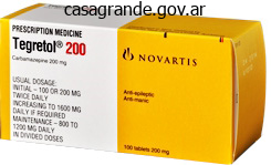
Tegretol dosages: 400 mg, 200 mg, 100 mg
Tegretol packs: 30 pills, 60 pills, 90 pills, 120 pills, 180 pills, 270 pills
Buy tegretol with paypal
The anterior half is the supramarginal gyrus, which arches over the upturned end of the lateral fissure. It is steady anteriorly with the lower part of the postcentral gyrus and posteroinferiorly with the superior temporal gyrus. The middle a half of the inferior parietal lobule, called the angular gyrus, arches over the tip of the superior temporal sulcus and is steady posteroinferiorly with the middle temporal gyrus. The posterior part of the inferior parietal lobule arches over the upturned end of the inferior temporal sulcus onto the occipital lobe, forming an arcus temporo-occipitalis. Area 3a lies most anteriorly, apposing area four, the first motor cortex of the frontal lobe; area 3b is buried in the posterior wall of the central sulcus; space 1 lies alongside the posterior lip of the central sulcus; and area 2 occupies the crown of the postcentral gyrus. The major somatosensory cortex contains within it a topographical map of the contralateral half of the physique. When seen acutely, the patient denies her motor deficit (anosognosia) and in fact denies the very existence of her paretic facet (hemisomatotopagnosia). With sensory testing, she ignores (neglects) the left facet of her body, as demonstrated with double simultaneous stimulation, and fails to attend to the left side of space. Discussion: this lady exhibits a severe disorder of the body scheme (body image) because of an acute (vascular) lesion (infarction) involving primarily the right (non-dominant) parietal cortex. Impaired proprioception and no much less than some degree of mental confusion are attribute elements of the medical syndrome. Within several days of onset, the hemisomatotopagnosia disappears, and she becomes increasingly aware of the existence of her hemiparesis. Some think about areas 22 and 37 to be auditory and visuo-auditory areas, respectively, related to speech and language. Within the ventral posterior nucleus, anteroposterior rods of cells respond with comparable modality and somatotopic properties. An apparently stepwise hierarchical progression of knowledge processing occurs from area 3b by way of space 1 to area 2. Callosally projecting pyramidal cells receive monosynaptic thalamic and commissural connections. Corticostriatal fibres, arising in layer V, move primarily to the putamen of the same facet. The topographical representation in the cortex is preserved by means of the spinal segments to which totally different elements of the postcentral gyrus project. Thus, the arm representation projects to the cervical enlargement, the leg representation to the lumbosacral enlargement and so forth. Fibres from areas 3b and 1 end extra dorsally, and people from space 2 extra ventrally. Superior and Inferior Parietal Lobules Posterior to the postcentral gyrus, the superior a half of the parietal lobe consists of areas 5, 7a and 7b. The thalamic afferents to this space come from the lateral posterior nucleus and from the central lateral nucleus of the intralaminar group. Ipsilateral corticocortical fibres from space 5 go to space 7, the premotor and supplementary motor cortices, the posterior cingulate gyrus and the insular granular cortex. Commissural connections between space 5 on each side are most likely to avoid the areas of illustration of the distal limbs. Connections pass to the posterior cingulate gyrus (area 23), insula and temporal cortex. Area 7b is reciprocally related with space forty six within the prefrontal cortex and the lateral part of the premotor cortex. Thalamic connections are with the medial pulvinar nucleus and the intralaminar paracentral nucleus. The major ipsilateral corticocortical connections to space 7a are derived from visible areas in the occipital and temporal lobes. In the ipsilateral hemisphere, area 7a has connections with the posterior cingulate cortex (area 24) and with areas eight and 46 of the frontal lobe. Area 7a is connected with the medial pulvinar and intralaminar paracentral nuclei of the thalamus.
Purchase 400 mg tegretol amex
A verify valve (2) permits controlled pressure by way of a needle (5) & into the shielded vial containing the Y (4). The strain pushes the suspension by way of an output needle (5), then through a 2nd heck valve (2), & lastly via the delivery catheter (6). This indicated aberrant vascular anatomy, arterioportal shunting, or overzealous injection. A selective arteriogram with a microcatheter within the left hepatic artery famous aberrant vascular anatomy. Conversely, the decrease branch vacillates distally-proximally, elevating considerations as the right gastric artery. Nontarget radioembolization of the best gastric artery may cause significant morbidity, even mortality. While not necessary, this large cystic artery was additionally embolized due to its size. Inadvertent radioembolization of this vessel might end in anterior stomach pain &/or skin/fat necrosis. Inadvertent radioembolization could end in duodenal ulceration and even perforation. Right Gastric Embolization through Left Gastric Artery (Arteriogram) Right Gastric Embolization through Left Gastric Artery (Post Coil Embolization) (Left) During a planning arteriogram, the proper gastric artery was seen arising off a bend at the bifurcation of the left hepatic & right hepatic arteries. Extrahepatic Tc99 activity ought to immediate additional investigation to prevent nontarget embolization. The large dimension (4 cm) and presence of pseudoaneurysms predispose to hemorrhagic issues. Restricted diffusion is seen in renal cell cancers and could also be helpful in differentiating these from benign lesions. The renal mass is separated from the liver with hydrodissection carried out prior to ablation. The location of this mass makes it an excellent candidate for percutaneous cryoablation. The patient was not a surgical candidate because of comorbidities, and the renal mass was an applicable size for ablation. A skinny rim (< 5 mm) of enhancement may sometimes be seen, which is a benign ancillary discovering. Note that the mass extends centrally and is in close proximity to the renal pelvis. There is risk of nontarget thermal damage during ablation, which may end in ureteral stricture &/or urinoma. Review of multiplanar reformats is crucial in assessing all ablation zone margins. In the absence of downstream obstruction within the renal collecting system, this is a benign ancillary finding. While true tumoral seeding of the tract is exceedingly rare, enlarging or persistent suspicious soft tissue might necessitate biopsy. Perirenal hemorrhage could be seen after ablation; rarely, blood loss is critical enough to require or extend hospital admission. The tines of the probe prolong beyond the border of the mass in axial (shown), coronal, and sagittal (not shown) planes. Unfortunately, on the 3-month medical follow-up, the affected person complained of arm weak spot (dropping espresso cups) and capturing arm pain. Fortunately, arm strength returned shortly thereafter, while ache resolved after ~ 9 months. As essential, the probe trajectory is corrected and aligned with the nodule prior to pleural entry. Minimizing the number of pleural punctures reduces the danger of procedure-related pneumothorax. A halo (> 5 mm) completely encircling the nodule is a predictor of remedy success.
Buy generic tegretol from india
The typical findings are of airway dilatation (usually defined as a diameter higher than that of the accompanying branch of the pulmonary artery). Temporary dilatation can happen throughout acute infections so the scan must be performed when the affected person is secure. The facilitation (or maximisation) of clearance of sputum and the therapy of infection with antibiotics and of bronchoconstriction with bronchodilators. Twicedaily postural drainage (20 minutes morning and night) or newer physio therapy techniques should be recommended. The use of antibiotics throughout exacerbations is routine, although of unproven profit, and prednisone in tapered doses may be useful. Patients with frequent infections could profit from longterm antibiotic treatment with macrolides. Recom binant deoxyribonuclease is useful for cystic fibrosis sufferers, however may be dangerous for different sufferers with bronchiectasis. It can pose a diagnostic and management downside, and remains a number one explanation for cancer dying in men and women. At diagnosis only 20% of patients could have local illness and half may have disseminated illness. Over the past 25 years, adeno carcinoma has become extra frequent than squamous cell carcinoma. Massive haemoptysis might occur because of bronchial wall erosion and the increased vascularity of the bronchial walls. Surgery must be thought-about for localised disease to stop development, or as therapy of intractable haemorrhage. Find out how the prognosis was made or suspected � a proportion of sufferers are asymptomatic and were diagnosed after a routine chest Xray or scan. Enquire about a history of unresolved pneumonia (a frequent reason for investiga tions to exclude carcinoma of the lung), pleural effusion or lung abscess. Hypercalcaemia (increased parathyroid hormone occurs often in squamous cell carcinoma) b. Hypertrophic pulmonary osteoarthropathy (may be extra common with adenocarcinoma) 4. Opportunistic infections (most typically in these handled with chemotherapy) ht tp:// eb oo ks m ed ebooksmedicine. Sputum cytology may be useful in centrally positioned lesions, however fibreoptic bronchos copy is now extra often done first. Bronchial brushings and washings, taken at bronchoscopy, also needs to be despatched for cytological examination but have a lower yield than biopsy. Chest indicators will vary � hear carefully for a set inspiratory wheeze over a big bronchus. Recurrent laryngeal nerve palsy may have caused hoarseness, and phrenic nerve paralysis may have triggered an elevation of a hemidiaphragm. It has been shown to detect metastases in as a lot as 20% of patients beforehand thought go properly with ready for surgery. The discovering of a solitary nodule on a chest Xray have to be investi gated, but benign causes embody a postinfection granuloma or a hamartoma. Other investigations could include bone marrow biopsy, mediastinoscopy and thora cotomy. Symptoms and signs that suggest central nervous system, liver, bone, chest wall or mediastinal involvement need to be sought fastidiously. Some nucleated pink cells (normoblasts) and myeloid cells (metamyelocytes and myelocytes). Comment: the mix of normochromic anaemia and the presence of marrow precursors in the peripheral blood is identified as a leukoerythroblastic response. The abnormal liver function exams are non-specific, but suggest liver involvement in this setting. In small cell carcinoma, stage the disease into restricted disease (lung primary, ipsi lateral and contralateral hilar, mediastinal and supraclavicular nodes) or in depth illness � 70% of patients (contralateral lung, distant metastases). In common, illness confined to one hemithorax and the ipsilateral cervical nodes known as restricted and further involvement is described as extensive illness. In patients with restricted illness, chemotherapy and concurrent radiotherapy enhance the prognosis.
Order tegretol 100mg on line
This is now usually a chronic illness and sufferers are getting older and more probably to have problems such as ischaemic coronary heart disease. The candidate might be expected to display a logical method to the case and, in fact, present a sympathetic attitude to the patient with this chronic illness. There needs to be a powerful emphasis within the dialogue on the psychological and social effects of the illness. If the patient is assumed to have a fever caused by a drug � a drug fever � it could be worth stopping the drug or utilizing another one. There is usually some combination of the following symptoms: � fever � vomiting � lymphadenopathy � diarrhoea � maculopapular rash � headache � arthralgia � meningism � myalgia � weight reduction � pharyngitis � oral candidiasis � nausea Ask concerning the mode of acquisition of an infection. Co-morbidity differs between subgroups, so specific questions on threat elements are important. Contact tracing have to be talked about and the risk of infection of sexual companions with out their knowledge addressed. The lesion is reddish-brown because of its vascular nature and accompanying haemosiderin deposition. Find out about antiretroviral drug and antibiotic treatment and about any adverse results of those. If the illness has lately been diagnosed, ask what remedy choices have been mentioned with the patient. Amprenavir Diarrhoea, nausea, perioral paraesthesia, lipodystrophy, insulin resistance ne Nelfinavir Diarrhoea, lipodystrophy, insulin resistance, hepatitis t/i Saquinavir Diarrhoea, lipodystrophy, insulin resistance, hepatitis nt er Indinavir Nausea, diarrhoea, lipodystrophy, insulin resistance, hepatitis, renal calculi, hair and nail adjustments na C. False positives can happen related to current vaccinations, other viral infections ht tp:// eb oo ks m ed ebooksmedicine. Other checks are used to assess the severity of the sickness and immunocompromise, and to search for related situations. The presently related checks will rely upon the scientific presentation; nevertheless, sure exams are important for all sufferers, either as a baseline or for monitoring (Table 13. Herpes simplex prophylaxis with acyclovir and Candida prophylaxis with fluconazole or ketoconazole are applicable for prevention after one episode has occurred. Drugs must be used solely in combination to ht tp:// eb oo ks m ed ebooksmedicine. Serial full blood counts and biochemistry will assist assess any complications of disease or therapy. Resistance testing is indicated for the remedy of the naive affected person, or if remedy is failing and shall be changed. Treat the mom with zidovudine as an infusion during labour, and treat the toddler for six weeks. Surveillance for the development of hepatic carcinoma can be indicated for these sufferers. Special care with drug monitoring is important if the affected person has cirrhosis; zidovudine and didanosine ought to be averted. Disseminated cytomegalovirus infection � retinitis, gastrointestinal illness m ed ebooksmedicine. Patients whose illness is nicely controlled by antiviral agents usually tend to die of heart problems than infection. Hyperlipidaemia ought to be treated with pravastatin (which has less interplay with antiviral drugs). If one of many examiners is a geriatrician then questions on this matter are very doubtless (from the opposite examiner). Measure the time it takes the patient (wearing their regular footwear and with any aides) to rise up from a chair, stroll 3 metres, turn round and stroll again, then sit. With the affected person back in bed, carry out cerebellar testing and look for peripheral neuropathy. Does the affected person undertake dangerous activities: climbing ladders, clearing gutters, and so forth.
Best buy for tegretol
Most human cerebellar disorders contain the anterior or posterior lobe, or each, or their outflows, inflicting the monitoring system to be lost and discovered movements to turn out to be clumsy. Many motor abilities require precise timing, which includes an excessive degree of cooperation between prime movers and their antagonists. For instance, studying a printed page requires that the scanning eyes snap again to the start of a line, time after time. Even small errors could end in dyslexia, whereby slight incoordination of eye movements causes the letters of a word to seem jumbled. Clinical testing of timing may be carried out simply by checking the flexibility to carry out rhythmic actions, similar to repetitive pronation� supination. Contraction of the ankle flexors causes the forefeet to push the decrease limbs and trunk backward in the meanwhile the hand grasps the guide. Once the carry gets under means, the erector spinae muscular tissues right for the combined weight of the guide and the reaching arm to forestall forward sway of the top and trunk. For example, throughout speech, the proper posterolateral area of the cerebellum is energetic bilaterally, which displays its function in coordinating the muscles concerned. Moreover, as a end result of proper lateral cerebellar exercise is even higher during practical naming. Examination demonstrates truncal ataxia, generally accompanied by incoordination of the limbs; variable ophthalmoparesis; and papilloedema on funduscopic examination. Discussion: Medulloblastoma usually presents with a midline cerebellar syndrome, with hydrocephalus and resultant increased intracranial stress. Clinically, it could be distinguished from ependymoma involving the fourth ventricle by the early appearance of nausea and vomiting in the latter, because of involvement of the fourth floor of the ventricle, including the realm postrema. Cranial nerve palsies may appear with either tumour, and rising intracranial strain is typical of each. The predominance of signs suggesting primary involvement of the vermis distinguishes medulloblastoma from cystic (or solid) astrocytoma of the cerebellum, which usually entails a cerebellar hemisphere rather than the vermis (although midline astrocytomas could cause diagnostic confusion). These tumours, which are highly delicate to radiotherapy, assault the pathway from the vermis to the nuclei of the vestibular nerves. The ataxia reflects malfunction of the linkage between the vermis and the lateral vestibular nucleus, which signifies that the antigravity assist normally pushed by the lateral vestibulospinal tract is lost or impaired. Scanning movements of the eye are inaccurate as a end result of the vermis no longer controls the gaze centres effectively. Disease of the anterior lobe is most often observed in continual alcoholics and presumably results from extended thiamine deficiency. Postmortem studies reveal pronounced shrinkage of the cortex of the anterior lobe. There could be losses of up to 10% of granule cells and 20% of Purkinje cells, and a 30% discount within the thickness of the molecular layer. The principal anatomical impact is atrophy of the connections between the anterior lobe and interposed nuclei and the reticulospinal pathways involved in normal locomotion. Incoordination of the decrease limbs results in a staggering gait and lack of ability to perform heel-to-toe walking. Anterior Lobe Lesions: Gait Ataxia Tendon reflexes could additionally be depressed in the lower limbs because of the loss of tonic stimulation of fusimotor neurones through the pontine reticulospinal tract. This causes a reduction of monosynaptic reflex activity throughout walking, which may finally produce stretching of sentimental tissues, a phenomenon that can end result in hyperextension of the knee joint throughout standing. Examination demonstrates a broad-based ataxic gait and ataxia with the heel�knee�shin check bilaterally. With the exception of signs of a light polyneuropathy, the remainder of the examination is normal. Discussion: the scientific features of a subacute evolving ataxia of the gait and of the legs, with good preservation of cerebellar operate within the upper extremities and little if another deficit, is typical of so-called alcoholic cerebellar degeneration occurring on a background of long-standing poor nutritional consumption. The comparatively restricted scientific syndrome, affecting primarily gait and the lower extremities, is defined by the observed distribution of lesions in the cerebellar cortex, involving predominantly the superior vermis and anterolateral portion of the cerebellar hemispheres-in accordance with identified somatotopic localization in the cerebellar cortex. All neurocellular parts of the cerebellar cortex could additionally be involved; Purkinje cells are most liable to damage.
Syndromes
- Bleeding from the umbilical cord just after birth
- Certain medicines (aspirin and other NSAIDS)
- Hematoma
- Grow very fast
- ERCP
- Wilson disease
- Hyperactivity
- Moderate to severe types of abnormal cell changes (called CIN II or CIN III)
Buy tegretol 100 mg
Resistance to one alkylating agent is commonly, but not all the time, related to resistance to the others. Relapse is handled for appropriate patients with autologous stem cell bone marrow transplant. Novel brokers corresponding to carfilzomib and pomalidomide are available by way of scientific trials or particular access applications. Two groups of patients are handled on this way: these with a malignant condition (Table 8. The options embody: Polyneuropathy Organomegaly Endocrinopathy Monoclonal gammopathy (osteosclerotic myeloma, or IgA or IgG M proteins with lambda light chains) Skin modifications. Treatment of the myeloma element within the traditional way often helps the opposite manifestations. The natural historical past is of progression of peripheral neuropathy to respiratory failure over some years. Find out whether or not there has been an issue leading to the current admission to hospital (if the affected person is an inpatient). Autoimmune disease (RhA, a quantity of sclerosis) ne t/i nt er na l-m vi de os 218 Examination Medicine are likely to have been made sterile by the remedy. Total physique irradiation and the use of alkylating agents are extra probably than different therapies to trigger everlasting sterility. Does the patient really feel higher or worse than before and what medium- and long-term prognosis has she or he been given These embrace skin changes similar to these of scleroderma, dry eyes and mouth (sicca syndrome), alopecia and bronchiolitis obliterans (signs of airflow obstruction). Examine for hepatic enlargement and ascites (veno-occlusive disease of the liver). Management after transplant begins with supportive therapy to forestall infection and bleeding earlier than engraftment occurs. Platelet restoration is often the slowest and tends to determine the time of discharge from hospital. It happens by definition within the first 3 months after transplant and is characterised by: � diarrhoea � skin rash � liver perform take a look at modifications. It is often handled with excessive doses of prednisone, antithymocyte globulin and monoclonal antibodies to T cells. Patients develop an autoimmune-like illness with sicca syndrome, arthritis, cholestasis and bronchiolitis obliterans. Bacterial infection is most common in the course of the early interval earlier than the neutrophil count reaches regular levels. Fungal infection can turn into an issue within a week of transplant, can be difficult to deal with and clearance of infection turns into quicker upon neutrophil restoration. Prophylactic therapy with fluconazole, cotrimoxazole and ganciclovir is often used to stop these issues. Most recover, however in extreme circumstances remedy with tissue plasminogen activator may be indicated. Overall, autologous bone marrow transplant is related to related but much less severe issues. Patients with non-malignant conditions can usually be very successfully handled with bone marrow transplant. For example, aplastic anaemia sufferers can count on as much as 90% disease-free survival. Opinions differ about the usage of bone marrow transplant as preliminary remedy for these patients. Ogden Nash (1902�71) Gait: get the affected person to walk to the end of the room, flip round and are available back. Note the size of stride, smoothness of stroll and turning around, stance, heel strike and arm swing. Hemiplegic, Parkinsonian, foot drop and other neurological gaits must be apparent. From behind � look at the backbone for scoliosis, muscle bulk of the shoulders, paraspinal muscular tissues, gluteal muscular tissues and calves; the iliac crests for lack of symmetry.
Discount 200 mg tegretol visa
It enters the inferior cerebellar peduncle, gives collaterals to the cerebellar nuclei and terminates, mainly ipsilaterally, in the vermis and adjoining areas of the anterior lobe and within the pyramis and adjoining lobules of the posterior lobe. The posterior thoracic nucleus receives primary afferents of every kind from the muscular tissues and joints of the lower limbs, which reach the nucleus through the gracile fasciculus. Accordingly, the tract transmits proprioceptive and exteroceptive details about the ipsilateral decrease limbs. Very quick conduction is required to maintain the cerebellum informed about ongoing actions. The axons within the posterior spinocerebellar tract are the biggest in the central nervous system, measuring 20 �m in exterior diameter. The upper limb equivalent of the posterior spinocerebellar tract is the cuneocerebellar tract. It informs the cerebellum about the state of activity of spinal reflex arcs related to the decrease limb and lower trunk. Its fibres originate in the intermediate gray matter of the lumbar and sacral segments of the spinal cord. They cross close to their origin and ascend close to the surface as far as the decrease midbrain before looping down within the superior cerebellar peduncle. Most fibres cross once more within the cerebellar commissure; thus, their distributions to the cerebellar nuclei and cortex appear to be the same as those of the posterior tract. The rostral spinocerebellar tract originates from cell groups of the intermediate zone and horn of the cervical enlargement. Although thought of to be the higher limb and higher trunk counterpart of the anterior spinocerebellar tract, most of its fibres remain ipsilateral throughout their course. It enters the inferior cerebellar peduncle and terminates in the same cerebellar nuclei and folia because the cuneocerebellar tract. The cuneocerebellar tract incorporates exteroceptive and proprioceptive elements that originate from the cuneate and external cuneate nuclei, respectively. The tract itself is predominantly uncrossed and ends within the posterior half of the anterior lobe. Exteroceptive and proprioceptive mossy fibre elements of the tract terminate differentially in the apical and basal a half of the folia. The exteroceptive part overlaps the pontocerebellar mossy fibre projection in the apices of the folia of the anterior lobe. Comparable units of ipsilateral proprioceptive and interceptive cerebellar projections exist for the intensive territory of the trigeminal mind stem nuclei. These nuclei also project to the ipsilateral inferior olive, relaying there to the contralateral cerebellar cortex and deep nuclei. Localization within the Olivocerebellar System: Zones and Microzones - Climbing fibres originate exclusively from the contralateral inferior olivary advanced. Projections from the totally different subnuclei of the inferior olive terminate as climbing fibres on longitudinal strips of Purkinje cells within the cerebellar cortex. Collaterals finish on the cerebellar or vestibular goal nuclei of those Purkinje cells. A longitudinal zonal association is therefore attribute of the group of the olivocerebellar projection. Moreover, the olivocerebellar projection zones correspond exactly to the corticonuclear projection zones already described. Climbing fibres from the inferior olive are able to modify the cerebellar output in such a means that cells within every subnucleus of the inferior olivary complex monitor the output of a single cerebellar module. The inferior olivary complicated and its climbing fibres can be activated by tactile, proprioceptive, visual and vestibular stimulation and from the sensory, motor and visual cortices and their mind stem relays. A somatotopic association of physique parts, matching the olivary projections on to the cerebellar cortex, has been detected in animal experiments. Olivocerebellar Climbing Fibre Connections - the inferior olivary complicated may be subdivided right into a convoluted principal olivary nucleus and posterior and medial accent olivary nuclei. Olivary fibres type the olivocerebellar projection to the contralateral cerebellar cortex and give off collaterals to the lateral vestibular nucleus and to the cerebellar nuclei. Topographic distribution of olivary and corticonuclear fibers within the cerebellum: a review. The zonal patterns of the olivocerebellar and Purkinje�nuclear projections correspond precisely. The caudal halves of the posterior and medial accessory nuclei innervate the vermis.

Order tegretol 200mg visa
Focal, nodular, or irregular gallbladder thickening should raise suspicion for malignancy. There is an vague border with the liver, initially thought to be suspicious for carcinoma, but found to represent xanthogranulomatous cholecystitis at resection. The association between porcelain gallbladder and gallbladder most cancers is now thought of debatable. The management of gallbladder polyps relies on measurement, with polyps measuring 10 mm, typically requiring cholecystectomy. There is asymmetric wall thickening with intramural fluid collections and focal thinning of the wall at the fundus. There is a transition zone (waisting) within the physique with regular wall thickness within the neck. The gallbladder wall was thick and indistinct with loss of echogenicity at its interface with the liver. The gallbladder wall is thick and hypodense, with infiltration of the adjoining liver. Gallbladder carcinoma update: multimodality imaging analysis, staging, and treatment choices. Although subtle, sludge (which is lower in density than stones but higher in density than bile) is seen layering above the stones. This is the wallecho-shadow sign, which differentiates a big gallstone from a porcelain gallbladder. Although the wall was not thick, the affected person was treated with percutaneous drainage. There is pericholecystic fluid and echogenic fat in this diabetic affected person with gangrenous cholecystitis. The lumen was full of necrotic adenocarcinoma with muscle invasion in the neck only. The gallbladder is distended with diffuse hypoechoic wall thickening in this patient with acute calculous cholecystitis. The affected person had a positive Murphy sign in preserving with acute calculous cholecystitis. There is now edema of the gallbladder wall and pericholecystic stranding from acute cholecystitis, which later perforated. This was largely necrotic adenocarcinoma with a small invasive tumor within the gallbladder neck. The gallbladder: unusual gallbladder conditions and, uncommon presentations of the frequent gallbladder pathological processes. Complicated cholecystitis: the complementary roles of sonography and computed tomography. The reason for mild biliary ductal dilatation was due to an obstructing stone within the frequent bile duct (not shown). Biliary ductal dilatation was seen both proximal in addition to distal to the lesion due to copious mucin production. Abnormalities of the Distal Common Bile Duct and Ampulla: Diagnostic Approach and Differential Diagnosis Using Multiplanar Reformations and 3D Imaging. Biliary intraductal papillary-mucinous neoplasm manifesting solely as dilatation of the hepatic lobar or segmental bile ducts: imaging options in six sufferers. Sonographic Murphy sign was positive, suitable with acute calculous cholecystitis. Note the irregularity and absent enhancement of the right lateral wall, suggesting gangrenous cholecystitis. Passive hepatic congestion in a 56-year-old man with a latest myocardial infarction. There have been no signs or symptoms of biliary obstruction, and these findings were attributed to her prior cholecystectomy. The affected person underwent a successful surgical procedure with enteric cyst drainage into the duodenum.
Purchase tegretol online pills
Ask the affected person to perform a practical process, such as undoing a button or writing one thing. At the tip, ask whether you might examine all the opposite joints which may be likely to be concerned and the opposite systems more doubtless to be affected. Reprinted with permission from Elsevier (The Lancet, 2008, vol no 371(9618):1114). Note the erosions of the metacarpal heads, decreased cartilage within the joint spaces and erosion of the ulnar styloid (arrow). Method Expose both knees and thighs totally and have the patient lie on his or her back. Fixed flexion deformity must be assessed; inspect the knee from the side (a house beneath the knee shall be visible). Examine for effusions � the patella tap (ballottement) is used to confirm a large effusion. The fluid from the suprapatellar bursa is pushed by the hand into the joint space by squeezing the lower a part of the quadriceps after which pushing the patella downwards with the fingers. In sufferers with a smaller effusion, urgent over the lateral knee compartment could produce a noticeable medial bulge on account of fluid displacement. Test flexion and extension passively and notice the range of motion and the presence or absence of crepitus. The lateral and medial collateral ligaments are examined by having the knee barely flexed, holding the leg with the right hand and arm, steadying the thigh with the left hand and transferring the leg laterally and medially. The cruciate ligaments are tested by steadying the foot along with your elbow and shifting the leg anteriorly and posteriorly with the opposite hand. Hold the lower leg and foot, flex and extend the knee while internally and externally rotating the tibia. The juxta-articular aspects of the tibia and femur appear sclerotic, but the bones are generally osteoporotic. As ordinary, glance across the room for walking aids and at the affected person for signs of systemic illness, rashes, and so on. Look on the pores and skin of the toes and toes (for scars, ulcers and rashes), and look for swelling, deformity and muscle losing. Deformities affecting the forefoot embody hallux valgus and clawing and crowding of the toes (in rheumatoid arthritis). Passive movement of the talar joints (dorsiflexion and plantarflexion) and subtalar joints (inversion and eversion) have to be assessed. The best way to look at the subtalar and midtarsal joints is to repair the os calcaneus and ankle joint with the left hand whereas inverting and everting the midfoot with the right. The midfoot (midtarsal joint) permits rotation of the forefoot on a fixed hindfoot. Examining the individual toes is useful in seronegative spondyloarthropathies (a sausage-like swelling of the toe is characteristic of psoriatic arthritis). Feel the Achilles tendon for nodules and palpate the inferior aspect of the heel for tenderness (plantar fasciitis). Patients with rheumatoid arthritis can have involvement of the cervical backbone, hips and shoulders, and X-rays of these can also be obtainable. There is generalised loss of joint areas, and early harmful modifications are present. The space of the junction of the forefoot and midfoot has numerous erosions, which is a standard finding. The preliminary inspection confirms that this is a case of ankylosing spondylitis (Table 16. Look for deformity, inspecting from each the again and the facet, particularly looking for kyphosis and lack of lumbar lordosis. Before asking the affected person to lie down, check for active sacroiliac disease by springing the anterior superior iliac spines. A simple (and unreliable) test for sacroiliac disease is to push with the heel of the hand on the sacrum and note the presence of tenderness in both sacroiliac joint on springing. Examine the heels for Achilles tendinitis and plantar fasciitis, which are characteristic of the spondyloarthropathies. Examine the chest for decreased lung growth (chest enlargement of less than three cm on the nipple line suggests early costovertebral involvement) and for indicators of apical fibrosis.
Buy tegretol 200 mg fast delivery
Treatment: Surgical treatment is generally indicated as a result of the potential for permanent nasal deformity. An open fracture requires instant surgical care accompanied by tetanus prophylaxis or a tetanus booster. If the fracture is displaced and closed, it can be safely lowered in the course of the initial week after the harm. The displaced or depressed bone fragments may be lowered manually or with the help of a particular instrument (elevator). After the reduction, the nasal cavities should be packed to provide "inside splinting," and a plaster cast is applied externally. Affected constructions of the bony facial skeleton are the maxillary sinus, orbit, and the zygoma or zygomatic arch. An isolated fracture of the orbital flooring with a partial herniation of the orbital contents into the maxillary sinus is a particular type of lateral midfacial fracture known as a blow-out fracture. Symptoms: A depressed fracture of the zygoma presents clinically with facial asymmetry. Fractures of the orbital floor can cause diplopia on upward gaze as a end result of entrapment of the inferior rectus muscle. Sensory disturbances involving the cheek, ipsilateral higher lip, and lateral nasal wall recommend a direct or indirect fracture-induced lesion of the infraorbital nerve, which enters the buccal delicate tissues under the infraorbital margin and is commonly involved by fractures of the orbital floor. Diagnosis: Inspection: Swelling is often present as a outcome of subcutaneous hemorrhage (periorbital or "monocle" hematoma. Asymmetry of the affected facial half is most probably to occur with despair of the zygoma or zygomatic arch, depending on the situation and extent of the fracture. Enophthalmos signifies involvement of the orbital ground (herniation of orbital contents). The proper cheek is flattened relative to the other aspect because of a depressed fracture of the zygoma. Palpation: Concomitant soft-tissue swelling could make it difficult or inconceivable to palpate websites of bony discontinuity or displacement. The following areas ought to be examined: � Frontozygomatic suture (upper part of the lateral orbital rim) � Infraorbital margin (anterior bony margin of the orbital floor) � Zygomatic arch (often troublesome to evaluate as a end result of soft-tissue swelling) Sensory testing: Wisps of cotton can be utilized to test sensory operate on the wholesome and affected sides. Radiographs: Whenever a lateral midfacial fracture is suspected, standard sinus radiographs ought to be obtained (occipitomental and occipitofrontal projections, see. The zygomatic arches could additionally be poorly visualized in normal projections, and so a "bucket handle" view should be added when a concomitant zygomatic arch fracture is suspected. Treatment consists of discount and fixation of the bone fragments utilizing miniplates, interosseous wiring, or both. Although fractures of the central midface and anterior cranium base (frontal skull base, rhinobase) are sepa- Classification: Central midfacial fractures: the Le Fort classification. Frontobasal fractures: these are bony accidents to the anterior skull base and adjacent paranasal sinuses (frontal and sphenoid sinuses, ethmoid labyrinth). The Escher classification distinguishes four forms of frontobasal fractures based mostly on the location and extent of the fracture strains. Severe craniocerebral trauma also can lead to vision loss attributable to ocular destruction or injury to the optic nerve (nerve contusion, rupture of the nerve in an intact sheath because of sagittal brain movement within the skull). Diplopia due to oculomotor palsy from harm to the third, fourth, or sixth cranial nerve is considerably rare and occurs provided that the fracture line runs through the cavernous sinus. Extensive injuries with sites of bone dehiscence can result in cerebral prolapse, with Ascending an infection can occur via the adjacent paranasal sinuses in frontobasal fractures and can result in life-threatening intracranial problems. Facial soft-tissue accidents are often intensive but might provide little information on possible related bony accidents at the stage of the skull base. The next step after nasal inspection is to examine the oral cavity and oropharynx. Otoscopy or otomicroscopy should also be carried out to exclude a concomitant petrous bone fracture.
References
- Gammie A, Kaper M, Dorrepaal C, et al: Signs and symptoms of detrusor underactivity: an analysis of clinical presentation and urodynamic tests from a large group of patients undergoing pressure flow studies, Eur Urol 69:361n369, 2016.
- Vijayalakshmi IB, Devananda NS, Chitra N. A patient with aneurysms of both aortic coronary sinuses of Valsalva obstructing both ventricular outflow tracts. Cardiol Young. 2009;19:537-9.
- Sofroniew MV. Astrocyte failure as a cause of CNS dysfunction. Mol Psychiatry 2000;5:230-2.
- American Society of Anesthesiologists Task Force on Management of the Difficult airway. Practice guidelines for management of the difficult airway: an updated report by the American Society of Anesthesiologists Task Force on Management of the Difficult Airway. Anesthesiology. 2003;98:1269-77.


