
Caleb P. Bupp, M.D.
- Department of Medical Genetics
- Spectrum Health System
- Grand Rapids, Michigan
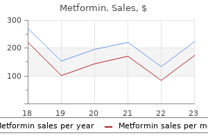
Metformin dosages: 850 mg, 500 mg
Metformin packs: 60 pills, 90 pills, 120 pills, 180 pills, 270 pills, 360 pills

Effective 500 mg metformin
Electron microscopy Electron microscopy exhibits electron-dense particles in phagolysosomes of macrophages. Cutaneous nodules containing globules of mercury have been reported in one patient, as a reaction to oral mercury. Other manifestations of persistent arsenical poisoning include keratoses, hyperkeratosis of the palms and soles, and carcinomas of the pores and skin. Periungual pigmentation developed in a number of the survivors of an incident involving the ingestion of arsenic-laced curry at a neighborhood pageant. The infiltrate around the follicles includes lymphocytes, plasma cells, and typically eosinophils. Macrophages with barely granular cytoplasm that stains purple-gray with H&E are often present. The aluminum can be confirmed by X-ray microanalysis846 or by the solochrome�azurine stain in which crystals of aluminum salts stain a deep gray-blue colour. Following the utilization of Drysol, there are individual and clusters of macrophages with ample cytoplasm. Pretibial pigmentation is more frequent, and that is slate grey to blue-black in color. Pigment granules � some staining for hemosiderin, some for melanin, and some for each � may be seen in macrophages and extracellularly. Histological examination showed the submucosal deposition of nice golden yellow granules, predominantly in a superficial location and in a linear fashion upon the extracellular matrix, particularly on elastic fibers. Phenothiazines Prolonged use of phenothiazines produces a progressive gray-blue pigmentation in sun-exposed areas. Electron microscopy shows melanin granules in macrophages but also different bodies of various electron densities that may represent metabolites or complexes of the drug. Refractile, golden-brown pigment is seen within the dermis, alongside collagen bundles, and in macrophages. The reported instances with deposits of minocycline pigment localized to the subcutaneous fat of the decrease extremity seem to be a unique kind (proposed kind V)887,888 A generalized muddy brown pigmentation due to increased melanin within the basal layer and accentuated in sun-exposed areas. Chlorpromazine and other phenothiazines produce slate-gray pigmentation in sun-exposed areas. The dermal pigment deposits related to these brokers can resemble melanin but may have a golden, refractile quality, possibly representing the drug or a metabolite thereof. Melanin stains are positive however, unlike minocycline pigmentation of the sort I variety, iron stains are adverse. The pretibial slate-gray pigment seen with antimalarial therapy can stain for iron, melanin, or both and due to this fact could be indistinguishable from minocycline deposition in the absence of a medical history or extra sophisticated laboratory research. The microscopic options of amiodarone and clofazimine pigmentation could probably be confused with each other or presumably with similar adjustments due to other drug metabolites. Background lesional modifications submitted for microscopic study would even be important; thus, a lesion of lepromatous leprosy or discoid lupus erythematosus containing these types of pigments would counsel clofazimine remedy. However, the purple deposits because of clofazimine, seen with fluorescence microscopy and in fresh frozen sections, seem to be characteristic, and laboratory strategies similar to vitality dispersive X-ray microanalysis and high-performance liquid chromatography might be used to identify amiodarone in tissue. Although usually believed to be lipofuscin, recent proof suggests that the material may in fact symbolize amiodarone itself. The pigment was originally thought to be lipofuscin, though melanincontaining complexes can also be present. Omeprazole Omeprazole, a potent inhibitor of gastric acid secretion, has been related to cutaneous pigmentation, mimicking ashy dermatosis. A biopsy revealed a traditional dermis and numerous macrophages containing golden-brown granules, mainly located around blood vessels within the higher dermis. The granules stained positively with the Masson� Fontana stain however had been adverse for iron. Sulfur-containing materials, representing the drug and/or drug�melanin complexes, was found in the cytoplasm of the macrophages. In the case of daunorubicin hyperpigmentation, full disappearance has been reported eight weeks after cessation of the remedy. A notuncommon side effect is the development of cutaneous and conjunctival pigmentation that has a reddish blue hue. An excellent evaluate of dermal filler supplies and botulinum toxin was revealed in 2001.
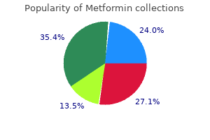
Discount metformin online master card
Solar lentigo, and even lentigo maligna, could additionally be related to degrees of keratinocyte atypia; equally, actinic keratoses may be accompanied by lentiginous modifications and/or junctional melanocytic hyperplasia. In such circumstances, a judgment should be made relating to which abnormal cell kind predominates. This can sometimes be aided by immunohistochemical staining for keratin, S100 protein, or significantly melan-A. Proliferation of atypical melanocytes along outer root sheaths of hair follicles favors lentigo maligna. Clinical knowledge can additionally be of help, although a spreading pigmented actinic keratosis can simply be confused with lentigo maligna, and in reality these two lesions can typically be juxtaposed. Exaggerated or broad-based basilar budding of actinic keratosis can occasionally be tough to distinguish from superficial basal cell carcinoma. Differentiation of actinic keratosis from types of porokeratosis can be a challenge because a cornoid lamella � the parakeratotic column that serves because the diagnostic hallmark for porokeratosis � can even occur in actinic keratosis, and levels of keratinocyte atypia are also observed in porokeratosis. However, keratinocyte atypia in porokeratosis is concentrated on the base of cornoid lamellae (representing an irregular clone of keratinocytes), whereas the atypia in actinic keratoses tends to be extra radially extensive and never confined to the neighborhood of the slim parakeratotic columns. On dermoscopy, the pigmented actinic keratosis has a putting similarity to lentigo maligna. It outcomes from chronic publicity to sunlight,558 though smoking and continual irritation may also contribute. Squamous cell carcinoma could develop after a latent period of 20�30 years,561,562 although the incidence of this transformation is troublesome to quantify. Treatment often parallels that given for actinic keratoses at different websites and contains cryotherapy, electrosurgery, imiquimod, retinoids, 800 Section7 � Tumors carbon dioxide laser ablation, and surgical excision. Other features are disordered maturation of epidermal cells, increased mitotic activity, and variable cytological atypia. These lesions have a superficial resemblance to the hyperkeratotic kind of seborrheic keratosis. The basal cell carcinomas that develop could also be of solid or (multifocal) superficial sort. Visceral cancers, significantly involving the lung and genitourinary system, can also be discovered. There is an increased risk of developing non-melanoma pores and skin most cancers, particularly with long-term, high-dose publicity. Several histological variants have been described, and multiple of those patterns may be present in numerous areas of the identical lesion. The verrucous�hyperkeratotic type is characterized by hyperkeratosis, papillomatosis, and sometimes intervening pit-like invaginations. Little has been printed in the dermatological or pathological literature on this change. Rare instances have been reported of metastatic squamous cell carcinoma presenting as epidermotropic bowenoid lesions. In a casual trial, dermatopathologists have been reliably able to categorize the continuum of keratinocytic atypia with substantial concordance. Mucinous and sebaceous metaplasia characterize two different rare histological patterns. This may lead to therapy failure when superficial methods of destruction are used. Invasion could additionally be facilitated by the manufacturing of metalloproteinases that are involved within the destruction of basement membrane. Occasional apoptotic our bodies are present within the intercellular areas, whereas others have been phagocytosed by neighboring keratinocytes. Both lesions could show aneuploidy of the constituent cells,749 and both may express mutant p53 protein and p21. Bowenoid atypia, including apoptotic keratinocytes and mitotic figures, can be seen in verrucae which were handled with podophyllin, although these adjustments largely resolve by seventy two hours following utility. These tumors represent an necessary public health problem,787�789 despite their comparatively low mortality rate.
Buy metformin 500mg otc
A key to the recognition of actinic keratosis is keratinocyte atypia that involves the basilar layer of the epidermis. Benign lichenoid keratoses can intently mimic actinic keratoses and might show limited degrees of atypia, at times making distinction from lichenoid actinic keratoses tough. However, the basilar atypia in benign lichenoid keratoses is often not solely mild but additionally confined to areas of most intense irritation. Evidence for origin in a lentigo or seborrheic keratosis is also frequently observed in benign lichenoid keratosis. Lichenoid actinic keratoses normally present unequivocal basilar atypia, usually extending past foci of band-like irritation, and suprabasilar acantholysis may be current. Forms of lupus erythematosus have been included within the differential prognosis of actinic keratoses, significantly the atrophic selection. The difficulties in distinguishing hypertrophic actinic keratoses from some irritated seborrheic keratoses (basosquamous acanthomas), especially those that occur in elderly individuals, were mentioned beforehand. Careful consideration to cytologic detail is important, notably with regard to basilar keratinocyte atypia, the presence of which favors hypertrophic actinic keratosis. However, occasional circumstances are tough to categorize, notably in superficial biopsy specimens, and re-excision may be warranted in these circumstances. Of these cases, 89 have been caused by squamous cell carcinoma, 22 by Merkel cell carcinoma, and 4 had been adenosquamous carcinomas. Clinicalaspects Basal cell carcinomas are discovered predominantly on areas of pores and skin exposed to the sun, notably in fair-skinned people. There are now calls for the use of supplemental vitamin D to counteract declining ranges of this vitamin in sun-protected people. Approximately 50% of all basal cell carcinomas studied have mutations of this gene. Squamous cell carcinomas are elevated greater than basal cell carcinomas, however the ratio of the two tumors nonetheless favors basal cell carcinoma. Mohs surgery is the treatment of alternative for primary facial basal cell carcinomas with aggressive histopathology and for recurrent tumors on the face. A evaluate of the chance elements, epidemiology, genetics, and treatment choices for basal cell carcinoma was published in 2011. Mutations in other genes in the sonic hedgehog pathway have also been found in sporadic tumors. They were preceded by publications stressing the significance of the histological sample. There is considerable variability in the morphology of basal cell carcinomas, and as a consequence, a quantity of histopathological subtypes have been outlined. Certain options are shared by more than one of these subtypes, and these elements are thought-about first. Basal cell carcinomas are composed of islands or nests of basaloid cells, with palisading of the cells on the periphery and a haphazard arrangement of these cells in the facilities of the islands. The tumor cells have a hyperchromatic nucleus with comparatively little, poorly outlined cytoplasm. There are quite a few mitotic figures, typically atypical,1058 and a correspondingly excessive number of apoptotic tumor cells. Lesions of long standing and aggressive tumors usually prolong into the decrease dermis. Deep extension happens both diffusely or throughout the paths of the cutaneous adnexae. Calcification may be present in the center of the keratin cysts that form in a number of of the histological subtypes. Similar basaloid proliferations could overlie a cutaneous myxoma1099 and connective tissue/ mesenchymal hamartomas. This is especially so with the (multifocal) superficial basal cell carcinoma the place nests can be extensively spaced or endure regression. It is sweet apply to order, routinely, three ranges of all punch and shave biopsies to forestall sampling errors. Clues in an preliminary nondiagnostic slide that suggest that deeper sections may yield basal cell carcinoma embrace focal basal atypia, stromal or superficial fibrosis, empty dermal spaces, equivocal adnexae, and microcalcifications.
Metformin 850mg line
Acne conglobata of the buttocks aggravated by mechanical and environmental components. Acne conglobata and a generalized lichen spinulosus-like eruption in a person seropositive for human immunodeficiency virus. Single nucleotide polymorphisms of toll-like receptor-4 shield towards pimples conglobata. Cutaneous side-effects in cancer sufferers, handled with the antiepidermal development factor receptor antibody C225. Necrotizing infundibular crystalline folliculitis manifesting as a perforating mucinosis: A case report. Epiluminescence dermatoscopy enhanced affected person compliance and achieved therapy success in pseudofolliculitis barbae. Pityrosporum folliculitis during pregnancy: A potential cause of pruritic folliculitis of being pregnant. Perforating folliculitis with jaundice in an Indian male: A uncommon case with sclerosing cholangitis. Perforating folliculitis related to tumour necrosis factor- inhibitors administered for rheumatoid arthritis. Perforating folliculitis in a affected person handled with nilotinib: A additional evidence of c-kit involvement. Sterile neutrophilic folliculitis with perifollicular vasculopathy: A distinctive cutaneous reaction sample reflecting systemic disease. Pseudolymphomatous folliculitis: A clinicopathologic research of 15 instances of cutaneous pseudolymphoma with follicular invasion. A evaluate of fifty five instances of cutaneous lymphoid hyperplasia: Reassessment of the histopathologic findings resulting in reclassification of four lesions as cutaneous marginal zone lymphoma and 19 as pseudolymphomatous folliculitis. Hyperplasia of hair follicles and different adnexal structures in cutaneous lymphoproliferative problems: A research of 53 cases, together with so-called pseudolymphomatous folliculitis and overt lymphomas. Light microscopic examination of scalp hair samples as an help in the diagnosis of paediatric issues: Retrospective evaluate of greater than 300 circumstances from a single centre. Human hair greying is linked to a particular depletion of hair follicle melanocytes affecting both the bulb and the outer root sheath. Trichotemnomania associated to trichotillomania: A case report with emphasis on the diagnostic worth of dermoscopy. Trichothiodystrophy: Review of sulfur-deficient brittle hair syndromes and association with the ectodermal dysplasias. Intractable diarrhea of infancy with facial dysmorphism, trichorrhexis nodosa, and cirrhosis. Characterization of tiger tail banding and hair shaft abnormalities in trichothiodystrophy. Trichothiodystrophy: Sulfur-deficient brittle hair as a marker for a neuroectodermal symptom complex. Trichothiodystrophy: Clinical spectrum, central nervous system imaging, and biochemical characterization of two siblings. Trichothiodystrophy related to photosensitivity, gonadal failure, and putting osteosclerosis. Cheveux incoiffables � Diagnostic, clinical and, hair microscopic findings, and pathogenic studies. Uncombable hair syndrome: Observations on response to biotin and incidence in siblings with ectodermal dysplasia. Pili canaliculi: Clinical and microscopic investigation of the first Brazilian family. Quantitative evaluation of scanning electron microscope defects in uncombable-hair syndrome. Straight hair related to interferon-alfa plus, ribavirin in hepatitis C infection.
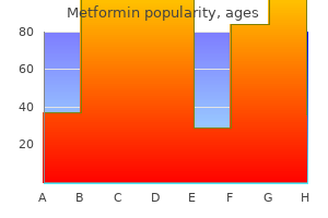
Metformin 850mg low cost
Efficacy of native warmth therapy by radiofrequency in the remedy of cutaneous leishmaniasis, compared with intralesional injection of meglumine antimoniate. Efficacy of a weekly cryotherapy regimen to deal with Leishmania major cutaneous leishmaniasis. Comparison between the efficacy of photodynamic remedy and topical paromomycin within the therapy of Old World cutaneous leishmaniasis: A placebo-controlled, randomized scientific trial. Interferon- remedy induces granulomatous tissue reaction in a case of localized cutaneous leishmaniasis. Treatment assessment by monitoring, parasite load in pores and skin biopsies from sufferers with cutaneous leishmaniasis, using quantitative nucleic acid sequence-based amplification. Clinical and histopathological options of zoonotic cutaneous leishmaniasis in Saudi Arabia. Clinical and histopathological traits of cutaneous Leishmaniasis in Sanliurfa City of Turkey including Syrian refugees. A quantitative study of epidermal Langerhans cells in cutaneous leishmaniasis brought on by Leishmania tropica. Unusual histopathological features of cutaneous leishmaniasis identified by polymerase chain response specific for Leishmania on paraffin-embedded pores and skin biopsies. Comparison of standard and polymerase chain reaction diagnostic techniques for leishmaniasis in the endemic region of Adana, Turkey. A case of mucosal leishmaniasis: Beneficial utilization of polymerase chain reaction for diagnosis. Cutaneous leishmaniasis reactivation 2 years after remedy brought on by systemic corticosteroids�First report. Chronic destructive ulcerative lesion of the midface and nasal cavity due to leishmaniasis contracted in Djibouti. Cutaneous and mucocutaneous leishmaniasis resembling borderline-tuberculoid leprosy: A new scientific presentation Late relapse of mucosal leishmaniasis of the tongue in a affected person after thymectomy. Unusual affiliation of American cutaneous leishmaniasis and acquired immunodeficiency syndrome. Disseminated mucocutaneous leishmaniasis ensuing from continual use of corticosteroid. A histological classification of mucocutaneous leishmaniasis in Brazil and its medical analysis. A easy and sensitive take a look at for field prognosis of, post kala-azar dermal leishmaniasis. Post-kala-azar dermal leishmaniasis occurring after 10 years of treated kala azar. Post-kala-azar dermal leishmaniasis in the Sudan: Clinical presentation and differential prognosis. Post-kala-azar dermal leishmaniasis with visceral leishmaniasis, or a rare presentation of visceral leishmaniasis with in depth pores and skin manifestations. Fine-needle sampling provides considerable diagnostic yield in lesions of submit kala azar dermal leishmaniasis: Analysis of four cases from north eastern India. Post-kala-azar dermal leishmaniasis: A light and electron microscopic research of 18 circumstances. Macroglossia as a manifestation of post-kala-azar dermal leishmaniasis: A case report. Postkala-azar dermal leishmaniasis coexisting with borderline tuberculoid leprosy. Atypical cutaneous histological options of visceral, leishmaniasis in acquired immunodeficiency syndrome. Visceral leishmaniasis with cutaneous lesions in a patient contaminated with human immunodeficiency virus. Post-kala-azar dermal leishmaniasis as an immune reconstitution inflammatory syndrome in a affected person with acquired immune deficiency syndrome. Pathology of post-kala-azar dermal leishmaniasis: A gentle microscopical, immunohistochemical, and ultrastructural study of pores and skin lesions and draining lymph nodes. Identification of parasite antigen, correlation of parasite density and irritation in pores and skin lesions of post kala-azar dermal leishmaniasis. Cutaneous leishmaniasis mimicking inflammatory and neoplastic processes: A scientific, histopathological and molecular research of 57 circumstances.
Syndromes
- Muscle spasms
- Fruity-smelling breath
- Nervousness
- Muscle aches and stiffness
- When traveling where contamination is more likely, eat only hot, freshly cooked food. Drink water only if it has been boiled. DO NOT eat raw vegetables or unpeeled fruit.
- Problems breathing
- Paranoia
- Problems with thinking or confusion caused by small strokes
- Pain in the penis
- Bluish color of the skin (cyanosis)

Metformin 500mg amex
It has been suggested that the neurofollicular hamartoma, trichodiscoma, and fibrofolliculoma are a half of the same spectrum of hamartomas489 and, as said beforehand, that the neurofollicular hamartoma should be recategorized as a spindle cell-predominant trichodiscoma. The trichodiscoma element is often a well-demarcated and non-encapsulated tumor, which regularly has a folliculosebaceous collarette of variable maturity. Neither molecular nor immunohistochemical methods distinguish pilomatrix carcinomas from pilomatricomas. It can unfold to regional lymph nodes531 and to visceral organs, significantly the lungs. The reported case was composed of small nests of glycogen-containing clear cells infiltrating the dermis and subcutis. The tumor is composed of pleomorphic basaloid cells with outstanding nucleoli and frequent mitoses. Proliferating pilomatricomas characteristic mostly basaloid cells with scant cytoplasm, hyperchromatic nuclei, and mitotic activity. However, these are symmetrical, wellcircumscribed lesions that demonstrate expansile development. However, the findings lend assist to the thought that pilomatrix carcinomas sometimes might indeed come up from preexisting pilomatricomas. Small samples of pilomatrix carcinomas might create confusion with lymphoepithelioma-like carcinoma539 or undifferentiated metastatic carcinoma. An underlying B-cell persistent lymphocytic leukemia was present in one case of trichoblastic carcinosarcoma and in addition in a trichogenic carcinoma in the identical collection. They are derived from the sebaceous gland, which begins its growth as a bulge or collar at the junction of the infundibulum and isthmus of the hair follicle. Maturation of the mantle occurs slowly in childhood with the accumulation of lipid in a few of the cells, forming sebocytes at the base of the mantle. Sebocytes enhance in number and size such that a fully developed sebaceous lobule is present by puberty. Mantles are best seen around vellus follicles on the face, but additionally they develop in affiliation with terminal follicles. Later in life, the sebaceous glands endure progressive involution so that mantles are again seen, this time as vestiges. Initially, Steffen and Ackerman instructed that sebaceous glands had a quantity of cycles of progress, involution, and relaxation, impartial of the cycle of the hair follicle. The synthesis and accumulation of lipids is a key step in the differentiation of sebaceous gland cells. The trichoblastic carcinoma (malignant trichoblastoma) is a high-grade carcinoma arising in a trichoblastoma. The tumor consisted of a multifocal proliferation of basaloid follicular cells with a retiform development sample surrounded by a stroma resembling the perifollicular sheath. They are stated to be restricted to the feminine breast, but the writer has seen a case from a male breast. The hamartomas are quite uncommon and include folliculosebaceous cystic hamartoma and steatocystoma. Organoid nevus (nevus sebaceus) includes other appendageal components and is taken into account with the complicated adnexal tumors (see p. They are mentioned to be organized along pores and skin pressure lines in the supra- and subclavicular area; they could be seen at different sites, such as the face588 and penis. Interestingly, sebaceous hyperplasia can be produced in rats by the topical application of citral (3,7-dimethyl-2,6-octadienal), a chemical used in foods as a flavoring agent. The sebocytes are smaller than traditional, and there are extra basal cells per unit basement membrane length than in normal glands. It is assumed to be associated to the method of dysplastic epithelial proliferation in transplant recipients and not to the consequences of cyclosporine (ciclosporin),580 although this has been disputed. It has additionally been seen in a bone marrow recipient; cyclosporine was implicated in this case. Sebaceous lobules and individual sebaceous cells are current within the lining epithelium. Schulz and Hartschuh presented convincing evidence that this lesion is a trichofolliculoma at its very late stage with the follicular constructions in a state of involution, comparable to the conventional hair cycle. The case reported with a outstanding neural component within the stroma has some features in widespread with the neurofollicular hamartoma (see p.
Buy metformin pills in toronto
The acanthotic kind is composed of broad columns or sheets of basaloid cells with intervening horn cysts. Apoptotic cells are current within the base of the lesion and in areas of squamous differentiation. A variant with intercellular mucin and small basaloid keratinoyctes with spindled cytoplasm has been known as an adamantinoid seborrheic keratosis. There are keratin-filled invaginations and corneal pseudocysts; melanophages and looped vessels within the dermal papillae that seem to be oriented obliquely are additionally found. Clusters of melanosomes, which are often membrane sure, may be discovered within the cells. In addition, scientific historical past is important as a end result of lesions of acrokeratosis verruciformis begin at delivery, during childhood, or at puberty, whereas seborrheic keratoses have a a lot later time of onset. The inverted follicular keratosis (basosquamous acanthoma of Lund) has an architectural resemblance to trichilemmoma, another lobulated, endophytic epithelial tumor that may be arranged about a central follicular construction. However, the latter tumors characteristic clear cells (due to the presence of glycogen), a distinctly palisaded basilar layer, and a surrounding cuticular basement membrane. Seborrheic keratosis-like lesions in children usually have a tendency to be both verrucae or epidermal nevi (discussed previously). Dermatopathologists regularly encounter hyperkeratotic lesions which might be extra squamoid than basaloid, lack horn cysts, and have varying degrees of irregular papillomatosis, acanthosis, or scale crust formation. This would appear to be a reasonable strategy within the absence of scientific information to the contrary. Of larger concern are seborrheic keratoses that mimic premalignant or malignant epidermal tumors. As noted previously, seborrheic keratosis with basal cell clear cells can resemble melanoma in situ however may be distinguished by the sample of nuclear displacement and notably by differential staining with cytokeratins and melanocytic markers. Special staining typically demonstrates sweat gland differentiation in these tumors. These lesions require particularly shut consideration to architectural and cytologic particulars. In distinction to squamous cell carcinoma, irritated seborrheic keratoses show limited pleomorphism, absence of atypical mitoses, and lack of great atypia in decrease epidermal layers and basilar keratinocytes. Most often, the diagnosis may be established with confidence using these tips, but there are occasional lesions that defy accurate classification. Melanoacanthoma has been recorded Histopathology341 Histological examination of the skin lesions has solely been made in isolated cases. In several cases, the sweat ducts have been dilated, and rarely they could be hyperplastic. Variable submucosal inflammation may additionally be noticed,383 and melanophages are readily identified. The lesion reported as a cystic clear cell acanthoma might symbolize this tissue reaction occurring in an epidermal cyst or dilated follicle. Lesions can carefully resemble psoriasis, but the sharp demarcation from adjacent epidermis and adnexal sparing are options not encountered in psoriasis. Seborrheic keratoses lack pale-appearing keratinocytes, possess pseudohorn cysts, and are sometimes hyperkeratotic and hypergranulotic. Eccrine poroma can have similar medical features, together with a location on the distal extremities, and microscopically, sharp demarcation of tumor from adjoining dermis can additionally be a attribute of poromas. However, in distinction to clear cell acanthoma, these lesions are usually composed of small, closeset, basaloid cells. The characteristic feature is the presence of clear cells scattered mainly among the basal cells, with a few cells in the malpighian layer. These changes could be delicate and simply missed; subsequently, scientific suspicion is usually necessary to permit definitive prognosis. Large cell acanthoma is assumed to comprise sunlight-induced clones of irregular cells, without a tendency to malignancy.
Purchase metformin with american express
Five years after this biopsy was taken, the affected person developed cutaneous lupus erythematosus. Because an inflammatory infiltrate may be present even in instances with no medical inflammatory features, this classification is outdated. A number of ocular and skeletal defects have been reported in people with anetoderma. They have been chronicled in a evaluate of the in depth European literature on this situation. However, in a single large sequence, a perivascular infiltrate of lymphocytes was present in all circumstances. Direct immunofluorescence in some cases of primary anetoderma exhibits a pattern of immune deposits similar to that of lupus erythematosus. Electron microscopy the elastic fibers that stay are fragmented and irregular in appearance, but the collagen is normal. Mutations in this gene are also liable for age-related macular degeneration, the main reason for irreversible visual loss within the Western world. This gene encodes the two subunit of the vesicular H+ pump, and the mutation causes impaired vesicular trafficking, tropoelastin secretion, and cell survival. A congenital disorder of glycosylation involving a defect within the biosynthesis of Nand O-glycans has additionally been found in patients with cutis laxa. A variable inflammatory reaction is current in the acquired cases with an related clinical inflammatory part. Abnormalities of collagen structure have been famous in a couple of reports520,523 but particularly excluded in others. However, in none of those circumstances do the remaining elastic fibers present the shortened contours and indistinct outlines characteristic of cutis laxa. Nevertheless, it has been noted that mild elastic tissue changes could also be tough to discern in biopsy specimens, even with traditional histochemical stains for elastic fibers. Antibody staining for molecules known to be concerned in cutis laxa will doubtless turn into the future diagnostic methodology of selection, no less than for figuring out particular types of the illness. Elastic fibers are shortened, range in diameter, and have indistinct, hazy borders. Despite a reasonable discount in elastin deposition in the skin, the scientific adjustments are relatively delicate with increased softness and mobility of the pores and skin. Morphometric analyses of elastic fibers have demonstrated a marked reduction in elastic fiber diameter and volume compared with healthy controls. In almost 50% of circumstances, erythema, urticaria, or burning precedes or coincides with the event of the lesions, suggesting that an inflammatory course of could additionally be concerned within the pathogenesis. Elastophagocytosis was present in one case, suggesting that this could be the mechanism for the loss of elastic fibers. Two cases of mid-dermal elastophagocytosis, presenting as persistent reticulate erythema, have been reported. Elastic tissue is usually preserved around appendages, even in the clinical subset with perifollicular involvement. Histopathology There are varied hair shaft abnormalities, together with pili torti, monilethrix, and trichorrhexis nodosa. Electron microscopy the elastic fibers in the reticular dermis present a paucity of the central amorphous component whereas retaining regular microfibrillary materials. Engulfment of elastic fibers by macrophages may be seen in circumstances which have histological evidence of elastophagocytosis. Neurodegenerative modifications, vascular insufficiency, Histopathology578 There is a discount in dermal elastic tissue. Reduced numbers of elastic fibers, not essentially resulting in scientific manifestations, may happen in the course of several other granulomatous disorders. Multinucleate large cells and macrophages seem to be answerable for the digestion of the elastic fibers. Histopathology Loss and fragmentation of elastic fibers and decreased collagen have been famous in one report of this condition. It appears to end result from degradation of existing fibers and never from the synthesis of new elastic material.
Discount metformin 500mg on-line
This condition is not regarded as a variant of eosinophilic folliculitis as a end result of interfollicular irritation is typically the predominant function. Eosinophilic pustulosis appears to be an appropriate 464 Section4 � TheDermisandSubcutis designation for these cases. Ziemer and B�er have questioned the existence of this variant, stating that beforehand reported instances embody a spectrum of eosinophil-rich dermatoses including scabies and different bite reactions. Drugs corresponding to allopurinol, carbamazepine, and minocycline have been associated with an eosinophilic folliculitis. Most follicles are preserved, but some present disruption or destruction of the wall by the inflammatory infiltrate. A recently reported case showed that the intraepidermal pustules involving the palm had been centered around the intraepidermal parts of eccrine ducts. Immunohistochemical staining detected dermcidin throughout the pustules � a peptide with antimicrobial properties secreted by the eccrine equipment. The different well-known eosinophilic folliculitis occurs in erythema toxicum neonatorum, however that situation occurs in the course of the first few days of life and presents as patchy erythema of the face, trunk, and proximal extremities and sometimes resolves inside 1 week. Because eosinophilic folliculitis can even result from dermatophytosis parasitic illness and certain drugs, scientific historical past, evaluation of medications, and particular stains for organisms could also be indicated in some circumstances. The coexistence of eosinophilic folliculitis and follicular mucinosis creates a possible diagnostic dilemma as a result of eosinophils are generally found in cases that in any other case current clinically and histopathologically as follicular mucinosis; in such instances, these cells are often in the minority. Careful attention should then be paid to the lymphocytes in the dermal and perifollicular infiltrates. A variable mononuclear cell infiltrate normally surrounds the higher dermal portion of the hair follicle. A folliculitis is an uncommon presentation of secondary syphilis and of a nematode infestation. The lesion begins as a painful, follicular papule with surrounding erythema and induration. A carbuncle is a coalescence of a quantity of furuncles that will result in multiple points of drainage on the skin surface. The cord is most probably the results of fibroblastic proliferation round a lymphatic vessel � a situation most frequently related to axillary surgical procedure in girls with breast cancer. It presents as an erythematous follicular eruption that might be maculopapular, vesicular, pustular, or polymorphous. This is often destroyed, although a residual hair shaft is usually current in the center of the abscess. The overlying dermis is eventually destroyed, and the surface is roofed by an inflammatory crust. Attempts to reveal organisms in standard histological preparations are often unsuccessful. The most constant finding in herpes folliculitis is lymphocytic folliculitis and perifolliculitis. The inner root sheath cells cornify abruptly, with out formation of a granular cell layer. Various organisms may be involved, particularly Trichophyton tonsurans, Microsporum canis, and M. Histopathology There is variable irritation of the follicle and perifollicular dermis. If disruption of the hair follicle occurs, a quantity of foreign body big cells may be current. Hyphae and arthrospores may be found within the hair shaft or on the floor, relying on the character of the an infection. Abscess formation with partial or full destruction of hair follicles happens in a kerion. The small oval yeast accountable could be seen inside the infected follicle and could also be discovered within the adjacent dermis following rupture of the follicle.
Metformin 850mg fast delivery
Centrally, there may be foci of epidermal keratinization and occasionally small squamous eddies. There is a peripheral layer of columnar cells with nuclear palisading resembling the outer root sheath of hair follicles. A case of desmoplastic trichilemmoma arising in an organoid nevus has been reported. However, a variant of basal cell carcinoma has been described with thickened basement membrane, able to mimicking trichilemmoma and different benign tumors. However, the inverted follicular keratosis has a closer resemblance to the irritated seborrheic keratosis and is commonly thought-about an endophytic variant of that lesion. Desmoplastic trichilemmomas can mimic invasive carcinomas due to the interdigitation of islands of epithelial cells with fibromyxoid connective tissue. These embrace multiple trichilemmomas,104 which are normally on the face, acral keratoses, palmar pits, and mucocutaneous papillomatous papules. Symptomatic tumors have been treated with salicylic acid, etretinate, and cryotherapy, with only slight improvement. There is a plate-like fenestrated subepidermal tumor composed of pale or pink-staining glycogen-containing cells, with a peripheral palisade of basal cells. Hair follicles getting into the tumor from beneath lose their identity and merge with it. The tumor of the follicular infundibulum seems to be the identical as that reported as a basal cell hamartoma with follicular differentiation. The illustrations within the report of a tumor known as an infundibular keratosis confirmed some options of the a number of infundibular tumor referred to beforehand and others of an inverted follicular keratosis (see later) however without squamous eddies. Nonspecific verrucous acanthomas and lesions resembling digitate warts may be present. With progressive descent into the dermis, the tumor cells present outer root sheath differentiation characteristic of the infundibulum, isthmus, stem, and anagen bulb. A collision tumor of trichofolliculoma and basal cell carcinoma has been reported. The central follicle, which opens onto the surface, normally accommodates keratinous materials and typically vellus hairs. The follicles that branch off the central follicle may in flip give rise to secondary and even tertiary follicles. This complicated sample is quite different from the hair follicle nevus (discussed previously), which is composed of mature vellus follicles. Solitary trichoepitheliomas are discovered as skin-colored papules, with a predilection for the nose, higher lip, and cheeks. Most of the lesions reported as large solitary trichoepitheliomas are trichoblastomas. Many of the instances of malignant transformation reported within the older literature symbolize cases of the nevoid basal cell carcinoma syndrome and never trichoepitheliomas. They are composed of islands of uniform basaloid cells, generally exhibiting peripheral palisading. Rupture of these cysts with liberation of the keratinous debris ends in a small international body granuloma in the stroma. Foci of calcification are often current; amyloid is usually considered to be unusual, although it was present in 33% of instances in one examine. Aggregations of fibroblasts, representing abortive makes an attempt to kind papillary mesenchyme (papillary mesenchymal bodies), are attribute of trichoepithelioma. Desmoplastic trichoepitheliomas often current as asymptomatic solitary exhausting annular lesions with a raised border and a depressed center. The cells sometimes contain paranuclear glycogen, a feature not seen in basal cell carcinomas. Individual cells include tonofilaments and are linked to adjacent cells by desmosomes. In contrast to trichoepithelioma, basal cell carcinoma is more apt to feature a fibromyxoid stroma, clefting artifact separating tumor islands from this adjoining stroma, and apoptotic foci within the presence of melanin deposition.
References
- Thyberg J. Effects of nicotine on phenotypic modulation and initiation of DNA synthesis in cultured arterial smooth muscle cells. Virchows Arch B Cell Pathol Incl Mol Pathol 1986; 52:25.
- Duran AC, Angelini A, Frescura C, et al. Anomalous origin of the right coronary artery from the left aortic sinus and sudden infant death. Int J Cardiol. 1994;45(2):147-9.
- El Wakil A, Doghman M, Latre De Late P, Zambetti GP, Figueiredo BC, Lalli E (2011). Genetics and genomics of childhood adrenocortical tumors. Mol Cell Endocrinol 336: 169-173.
- Fasihuddin, Q., Hasan, A.T. Ureteroscopy (URS): an effective interventional and diagnostic modality. J Pak Med Assoc 2002;52:510-512.
- Moretti F, Uberti-Foppa C, Quiros-Roldan E, Fanti L, Lillo F, Lazzarin A. Esophagobronchial fistula caused by varicella zoster virus in a patient with AIDS: a unique case. J Clin Pathol 2002;55:397.
- Raj GV, Peterson AC, Toh KL, et al: Outcomes following revisions and secondary implantation of the artificial urinary sphincter, J Urol 173:1242n1245, 2005.
- Taub JM. Effects of habitual variations in napping on psychomotor performance, memory and subjective states. Int J Neurosci 1979;9(2):97-112.
- Reichardt KV, Berg P. Cloning and characterization of a cDNA encoding human galactose-1-phosphate uridyl transferase. Mol Biol Med 1988;5:107.